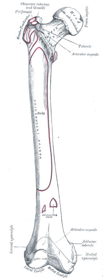
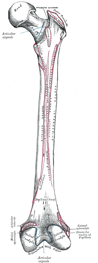
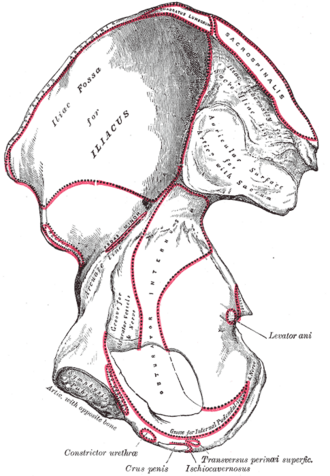
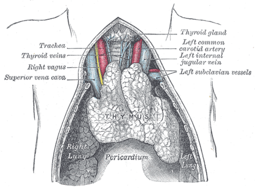
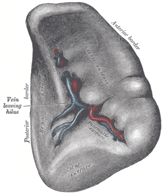
Image credits & licenses
This atlas only uses public-domain images, mainly plates from Gray's Anatomy of the Human Body (20th ed., 1918) hosted on Wikimedia Commons, plus original SVG schematics made for this project. The build rejects any image whose license isn't public domain or CC0.
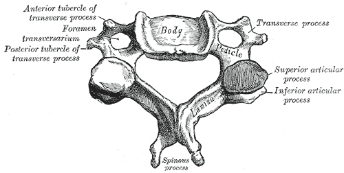 A cervical vertebra, from aboveHenry Vandyke Carter, Gray's Anatomy of the Human Body, 20th ed. (1918), Fig. 84. SourcePublic domain · Public domain in the US: published before January 1, 1931 (per Wikimedia Commons)Used in: Cervical vertebrae
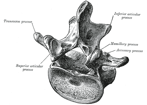 A lumbar vertebra from above and behindHenry Vandyke Carter, Gray's Anatomy of the Human Body, 20th ed. (1918), Fig. 93. SourcePublic domain · Public domain in the US: published before January 1, 1931 (per Wikimedia Commons)Used in: Lumbar vertebrae
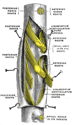 A portion of the spinal cord, right lateral surface, with the dura openedHenry Vandyke Carter, Gray's Anatomy of the Human Body, 20th ed. (1918), Fig. 796. SourcePublic domain · Public domain in the US: published before January 1, 1931 (per Wikimedia Commons)Used in: Spinal cord, Spinal meninges and spaces
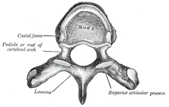 A typical thoracic vertebra, viewed from aboveHenry Vandyke Carter, Gray's Anatomy of the Human Body, 20th ed. (1918), Fig. 82. SourcePublic domain · Public domain in the US: published before January 1, 1931 (per Wikimedia Commons)Used in: Thoracic vertebrae
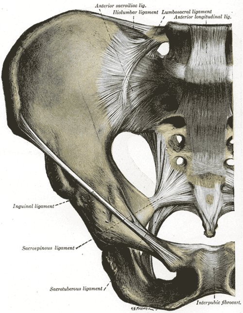 Anterior articulations of the pelvis (after Quain)Henry Vandyke Carter, Gray's Anatomy of the Human Body, 20th ed. (1918), Fig. 319. SourcePublic domain · Public domain in the US: published before January 1, 1931 (per Wikimedia Commons)
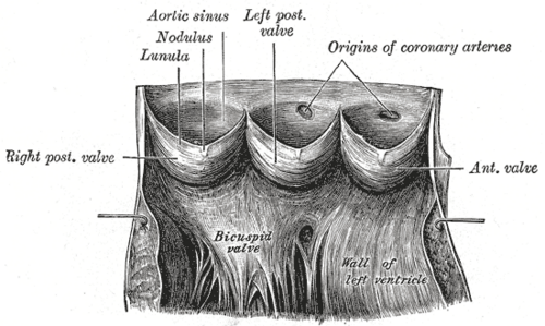 Aortic valve cusps and sinuses with the origins of the coronary arteriesHenry Vandyke Carter, Gray's Anatomy of the Human Body, 20th ed. (1918), Fig. 497. SourcePublic domain · Public domain in the US: published before January 1, 1931 (per Wikimedia Commons)
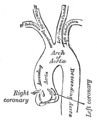 Arch of the aorta and its branchesHenry Vandyke Carter, Gray's Anatomy of the Human Body, 20th ed. (1918), Fig. 506. SourcePublic domain · Public domain in the US: published before January 1, 1931 (per Wikimedia Commons)Used in: Thoracic aorta
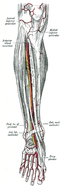 Arteries of the front of the legHenry Vandyke Carter, Gray's Anatomy of the Human Body, 20th ed. (1918), Fig. 553. SourcePublic domain · Public domain in the US: published before January 1, 1931 (per Wikimedia Commons)Used in: Popliteal artery
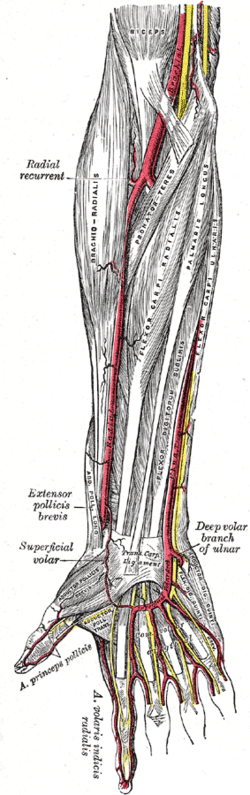 Arteries of the front of the right forearm and handHenry Vandyke Carter, Gray's Anatomy of the Human Body, 20th ed. (1918), Fig. 527. SourcePublic domain · Public domain in the US: published before January 1, 1931 (per Wikimedia Commons)Used in: Radial artery, Ulnar artery
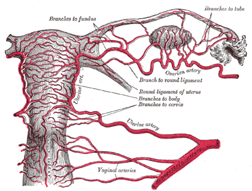 Arteries of the uterus and ovaryHenry Vandyke Carter, Gray's Anatomy of the Human Body, 20th ed. (1918), Fig. 1170. SourcePublic domain · Public domain in the US: published before January 1, 1931 (per Wikimedia Commons)Used in: Uterine artery, Uterus
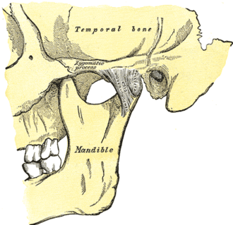 Articulation of the mandible, lateral aspectHenry Vandyke Carter, Gray's Anatomy of the Human Body, 20th ed. (1918), Fig. 309. SourcePublic domain · Public domain in the US: published before January 1, 1931 (per Wikimedia Commons)Used in: Temporomandibular joint
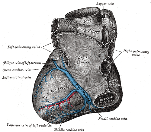 Base and diaphragmatic surface of the heartHenry Vandyke Carter, Gray's Anatomy of the Human Body, 20th ed. (1918), Fig. 491. SourcePublic domain · Public domain in the US: published before January 1, 1931 (per Wikimedia Commons)Used in: Circumflex artery
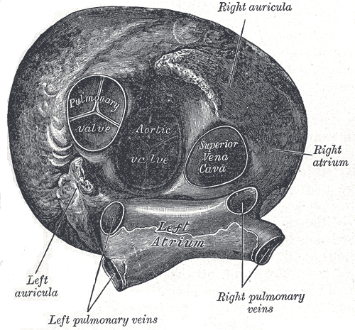 Base of the heart from above, showing the pulmonary and aortic valvesHenry Vandyke Carter, Gray's Anatomy of the Human Body, 20th ed. (1918), Fig. 494. SourcePublic domain · Public domain in the US: published before January 1, 1931 (per Wikimedia Commons)Used in: Pulmonary valve
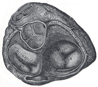 Base of the ventricles with the atria removedHenry Vandyke Carter, Gray's Anatomy of the Human Body, 20th ed. (1918), Fig. 495. SourcePublic domain · Public domain in the US: published before January 1, 1931 (per Wikimedia Commons)Used in: Mitral valve, Pulmonary valve
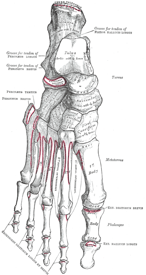 Bones of the right foot, dorsal surfaceHenry Vandyke Carter, Gray's Anatomy of the Human Body, 20th ed. (1918), Fig. 268. SourcePublic domain · Public domain in the US: published before January 1, 1931 (per Wikimedia Commons)Used in: Bones of the foot
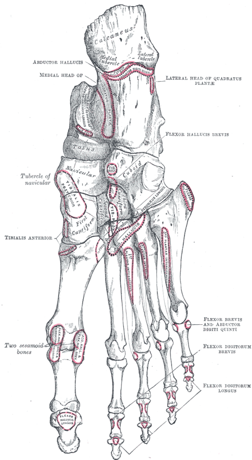 Bones of the right foot, plantar surfaceHenry Vandyke Carter, Gray's Anatomy of the Human Body, 20th ed. (1918), Fig. 269. SourcePublic domain · Public domain in the US: published before January 1, 1931 (per Wikimedia Commons)Used in: Bones of the foot
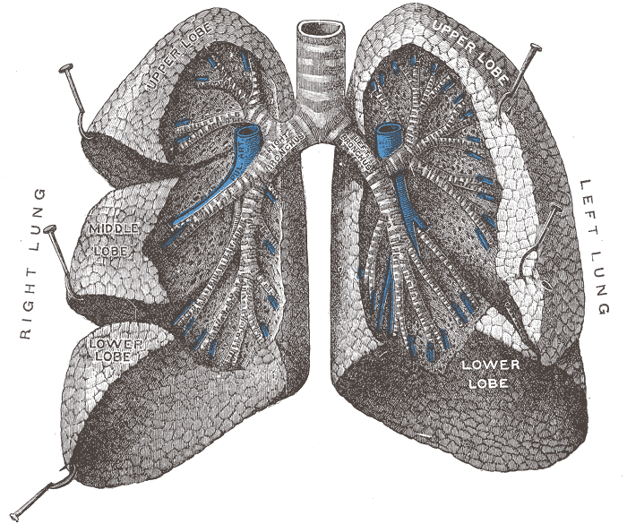 Bronchi and bronchiolesHenry Vandyke Carter, Gray's Anatomy of the Human Body, 20th ed. (1918), Fig. 962. SourcePublic domain · Public domain in the US: published before January 1, 1931 (per Wikimedia Commons)Used in: Lungs, Main bronchi
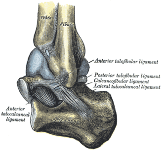 Capsule of the left ankle joint (distended), lateral aspectHenry Vandyke Carter, Gray's Anatomy of the Human Body, 20th ed. (1918), Fig. 356. SourcePublic domain · Public domain in the US: published before January 1, 1931 (per Wikimedia Commons)Used in: Lateral ankle ligaments
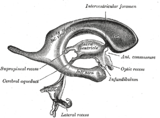 Cast of the ventricular cavities, side view (Retzius)Henry Vandyke Carter, Gray's Anatomy of the Human Body, 20th ed. (1918), Fig. 736. SourcePublic domain · Public domain in the US: published before January 1, 1931 (per Wikimedia Commons)Used in: Ventricular system and CSF
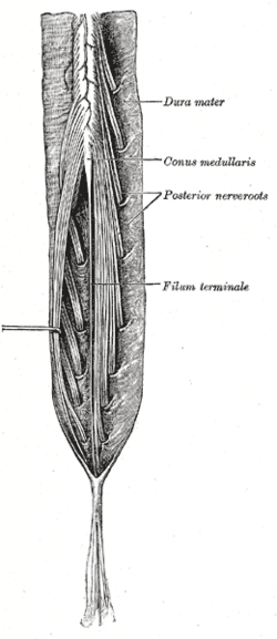 Cauda equina and filum terminale seen from behindHenry Vandyke Carter, Gray's Anatomy of the Human Body, 20th ed. (1918), Fig. 662. SourcePublic domain · Public domain in the US: published before January 1, 1931 (per Wikimedia Commons)Used in: Cauda equina
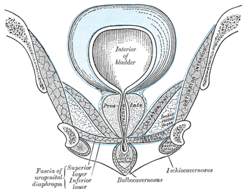 Coronal section of the anterior pelvis through the pubic archHenry Vandyke Carter, Gray's Anatomy of the Human Body, 20th ed. (1918), Fig. 407. SourcePublic domain · Public domain in the US: published before January 1, 1931 (per Wikimedia Commons)
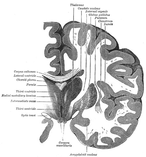
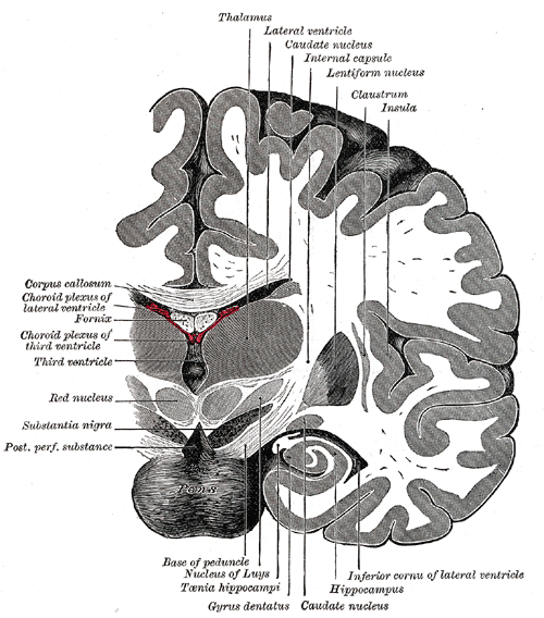
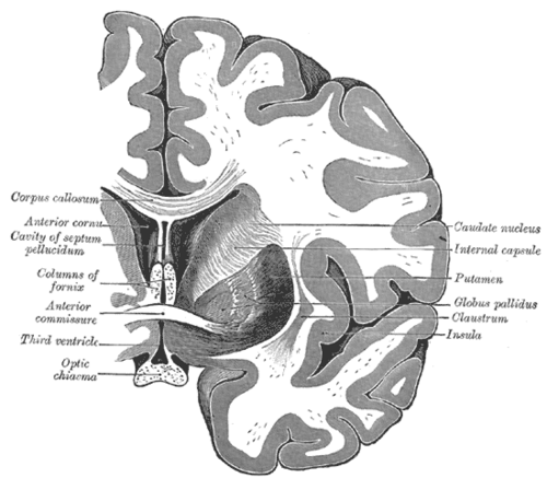 Coronal section of the brain through the anterior commissureHenry Vandyke Carter, Gray's Anatomy of the Human Body, 20th ed. (1918), Fig. 744. SourcePublic domain · Public domain in the US: published before January 1, 1931 (per Wikimedia Commons)Used in: Basal ganglia, Internal capsule
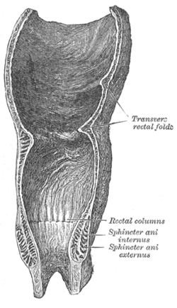 Coronal section of the rectum and anal canalHenry Vandyke Carter, Gray's Anatomy of the Human Body, 20th ed. (1918), Fig. 1078. SourcePublic domain · Public domain in the US: published before January 1, 1931 (per Wikimedia Commons)Used in: Anal canal
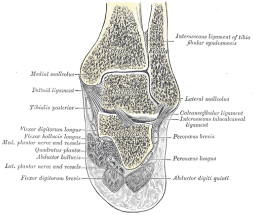 Coronal section through the right ankle and subtalar jointsHenry Vandyke Carter, Gray's Anatomy of the Human Body, 20th ed. (1918), Fig. 357. SourcePublic domain · Public domain in the US: published before January 1, 1931 (per Wikimedia Commons)Used in: Ankle joint
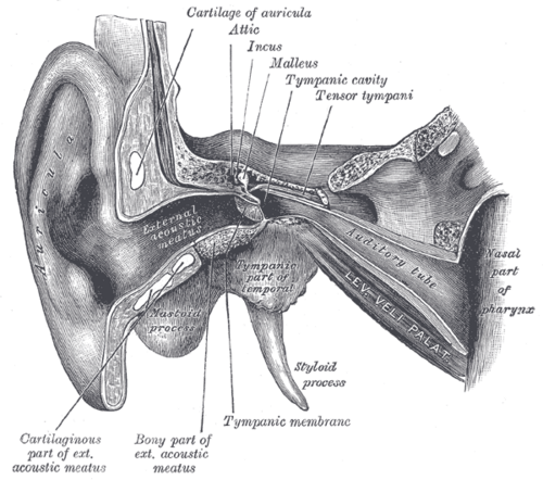 Coronal section through the right earHenry Vandyke Carter, Gray's Anatomy of the Human Body, 20th ed. (1918), Fig. 907. SourcePublic domain · Public domain in the US: published before January 1, 1931 (per Wikimedia Commons)
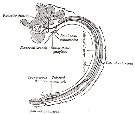 Course and branches of a typical intercostal nerveHenry Vandyke Carter, Gray's Anatomy of the Human Body, 20th ed. (1918), Fig. 819. SourcePublic domain · Public domain in the US: published before January 1, 1931 (per Wikimedia Commons)Used in: Intercostal space
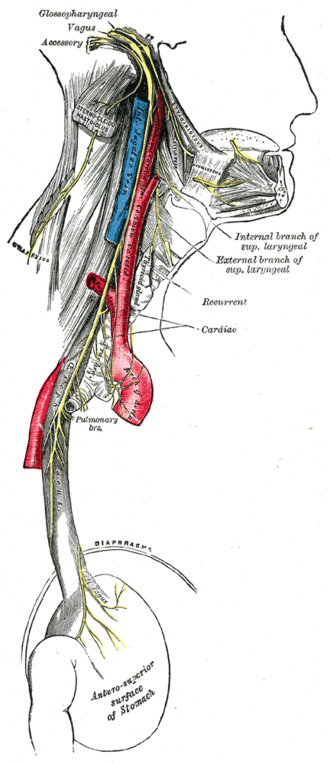 Course and distribution of the glossopharyngeal, vagus and accessory nervesHenry Vandyke Carter, Gray's Anatomy of the Human Body, 20th ed. (1918), Fig. 793. SourcePublic domain · Public domain in the US: published before January 1, 1931 (per Wikimedia Commons)
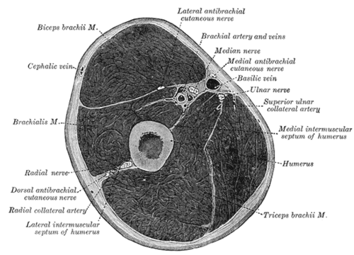 Cross-section through the middle of the armHenry Vandyke Carter, Gray's Anatomy of the Human Body, 20th ed. (1918), Fig. 413. SourcePublic domain · Public domain in the US: published before January 1, 1931 (per Wikimedia Commons)Used in: Biceps brachii, Ulnar nerve
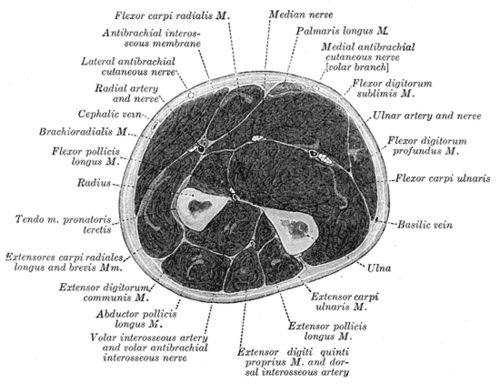 Cross-section through the middle of the forearmHenry Vandyke Carter, Gray's Anatomy of the Human Body, 20th ed. (1918), Fig. 417. SourcePublic domain · Public domain in the US: published before January 1, 1931 (per Wikimedia Commons)Used in: Median nerve
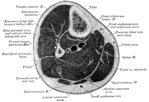 Cross-section through the middle of the legHenry Vandyke Carter, Gray's Anatomy of the Human Body, 20th ed. (1918), Fig. 440. SourcePublic domain · Public domain in the US: published before January 1, 1931 (per Wikimedia Commons)Used in: Compartments of the leg
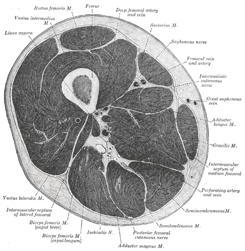 Cross-section through the middle of the thighHenry Vandyke Carter, Gray's Anatomy of the Human Body, 20th ed. (1918), Fig. 432. SourcePublic domain · Public domain in the US: published before January 1, 1931 (per Wikimedia Commons)Used in: Hamstrings, Quadriceps femoris
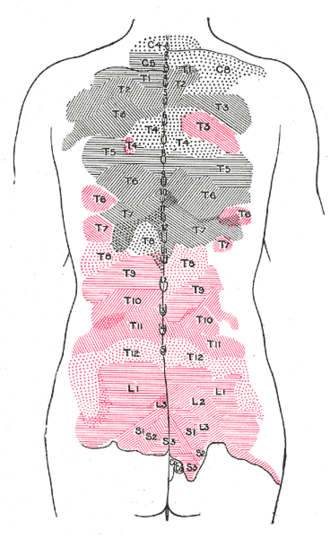 Cutaneous areas of the posterior divisions of the spinal nervesHenry Vandyke Carter, Gray's Anatomy of the Human Body, 20th ed. (1918), Fig. 802. SourcePublic domain · Public domain in the US: published before January 1, 1931 (per Wikimedia Commons)
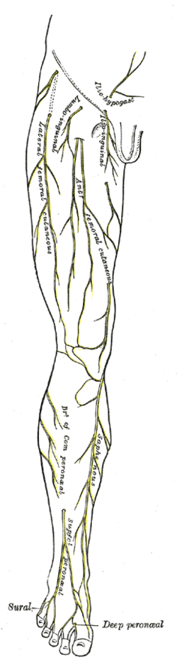 Cutaneous nerves of the right lower limb, front viewHenry Vandyke Carter, Gray's Anatomy of the Human Body, 20th ed. (1918), Fig. 825. SourcePublic domain · Public domain in the US: published before January 1, 1931 (per Wikimedia Commons)Used in: Femoral nerve
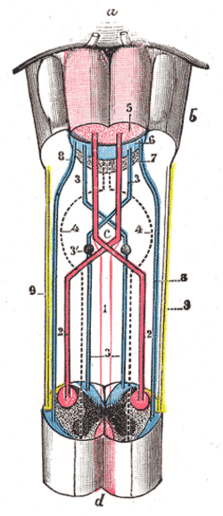 Decussation of the pyramids (scheme)Henry Vandyke Carter, Gray's Anatomy of the Human Body, 20th ed. (1918), Fig. 680. SourcePublic domain · Public domain in the US: published before January 1, 1931 (per Wikimedia Commons)Used in: Medulla oblongata
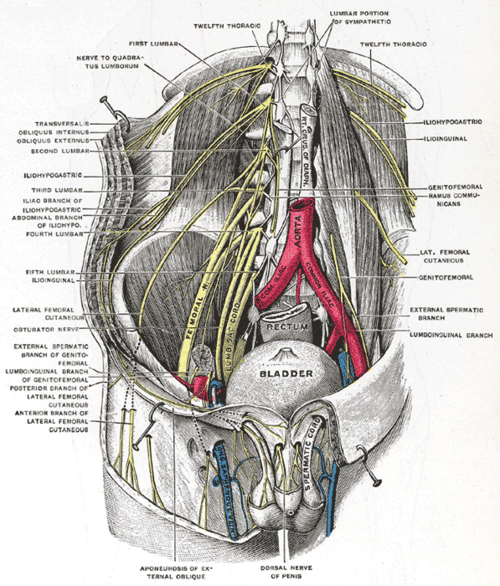 Deep and superficial dissection of the lumbar plexusHenry Vandyke Carter, Gray's Anatomy of the Human Body, 20th ed. (1918), Fig. 824. SourcePublic domain · Public domain in the US: published before January 1, 1931 (per Wikimedia Commons)Used in: Lumbar plexus, Psoas major
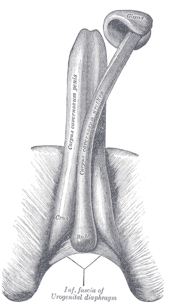
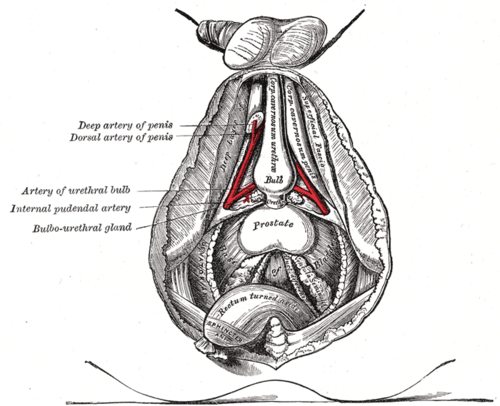 Deep dissection of the male perineumHenry Vandyke Carter, Gray's Anatomy of the Human Body, 20th ed. (1918), Fig. 543. SourcePublic domain · Public domain in the US: published before January 1, 1931 (per Wikimedia Commons)Used in: Perineal pouches
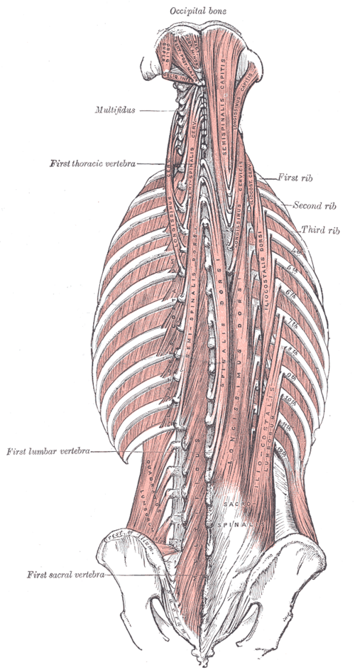 Deep muscles of the backHenry Vandyke Carter, Gray's Anatomy of the Human Body, 20th ed. (1918), Fig. 389. SourcePublic domain · Public domain in the US: published before January 1, 1931 (per Wikimedia Commons)Used in: Erector spinae
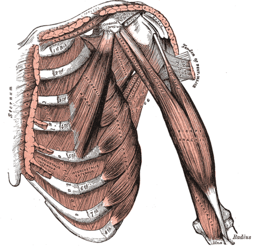 Deep muscles of the chest and front of the arm, with the boundaries of the axillaHenry Vandyke Carter, Gray's Anatomy of the Human Body, 20th ed. (1918), Fig. 411. SourcePublic domain · Public domain in the US: published before January 1, 1931 (per Wikimedia Commons)Used in: Subscapularis
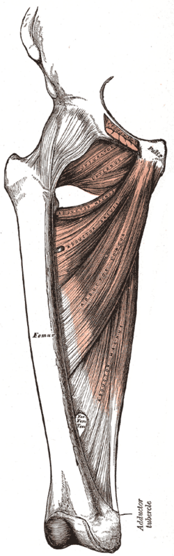 Deep muscles of the medial thighHenry Vandyke Carter, Gray's Anatomy of the Human Body, 20th ed. (1918), Fig. 433. SourcePublic domain · Public domain in the US: published before January 1, 1931 (per Wikimedia Commons)Used in: Adductor muscles of the thigh
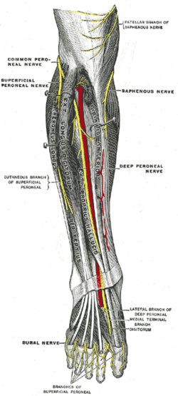 Deep nerves of the front of the leg (after Testut)Henry Vandyke Carter, Gray's Anatomy of the Human Body, 20th ed. (1918), Fig. 835. SourcePublic domain · Public domain in the US: published before January 1, 1931 (per Wikimedia Commons)Used in: Common fibular nerve
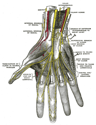 Deep palmar nerves (Testut)Henry Vandyke Carter, Gray's Anatomy of the Human Body, 20th ed. (1918), Fig. 817. SourcePublic domain · Public domain in the US: published before January 1, 1931 (per Wikimedia Commons)Used in: Ulnar nerve
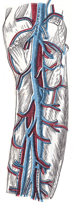 Deep veins of the back of the legHenry Vandyke Carter, Gray's Anatomy of the Human Body, 20th ed. (1918), Fig. 583. SourcePublic domain · Public domain in the US: published before January 1, 1931 (per Wikimedia Commons)Used in: Deep veins of the lower limb, Popliteal fossa
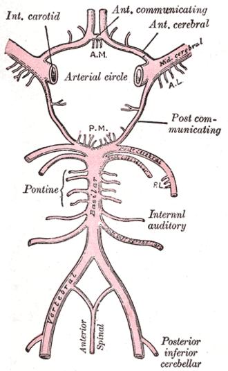 Diagram of the arterial circulation at the base of the brainHenry Vandyke Carter, Gray's Anatomy of the Human Body, 20th ed. (1918), Fig. 519. SourcePublic domain · Public domain in the US: published before January 1, 1931 (per Wikimedia Commons)Used in: Circle of Willis, Vertebrobasilar system
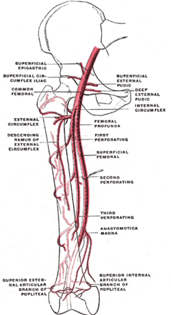 Diagram of the femoral artery and its branchesHenry Vandyke Carter, Gray's Anatomy of the Human Body, 20th ed. (1918), Fig. 548. SourcePublic domain · Public domain in the US: published before January 1, 1931 (per Wikimedia Commons)Used in: Femoral artery
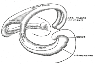 Diagram of the fornix (Spitzka)Henry Vandyke Carter, Gray's Anatomy of the Human Body, 20th ed. (1918), Fig. 747. SourcePublic domain · Public domain in the US: published before January 1, 1931 (per Wikimedia Commons)Used in: Limbic system
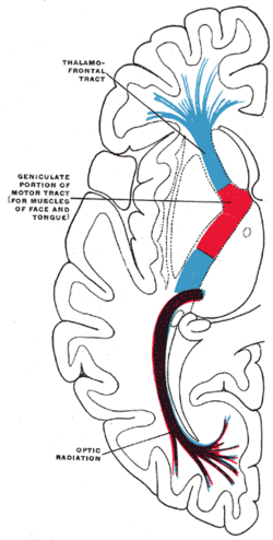 Diagram of the tracts in the internal capsuleHenry Vandyke Carter, Gray's Anatomy of the Human Body, 20th ed. (1918), Fig. 746. SourcePublic domain · Public domain in the US: published before January 1, 1931 (per Wikimedia Commons)Used in: Internal capsule
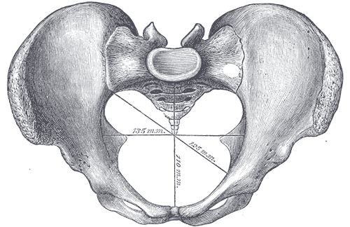 Diameters of the superior aperture of the lesser pelvis (female)Henry Vandyke Carter, Gray's Anatomy of the Human Body, 20th ed. (1918), Fig. 238. SourcePublic domain · Public domain in the US: published before January 1, 1931 (per Wikimedia Commons)Used in: Bony pelvis
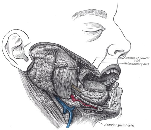 Dissection showing the salivary glands of the right sideHenry Vandyke Carter, Gray's Anatomy of the Human Body, 20th ed. (1918), Fig. 1024. SourcePublic domain · Public domain in the US: published before January 1, 1931 (per Wikimedia Commons)Used in: Parotid gland, Submandibular gland
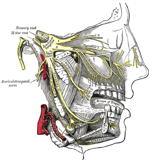 Distribution of the maxillary and mandibular nervesHenry Vandyke Carter, Gray's Anatomy of the Human Body, 20th ed. (1918), Fig. 778. SourcePublic domain · Public domain in the US: published before January 1, 1931 (per Wikimedia Commons)Used in: Trigeminal nerve (CN V)
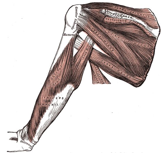 Dorsal muscles of the scapula and triceps brachiiHenry Vandyke Carter, Gray's Anatomy of the Human Body, 20th ed. (1918), Fig. 412. SourcePublic domain · Public domain in the US: published before January 1, 1931 (per Wikimedia Commons)
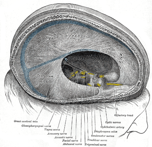 Dura mater and tentorium with the cranial nervesHenry Vandyke Carter, Gray's Anatomy of the Human Body, 20th ed. (1918), Fig. 567. SourcePublic domain · Public domain in the US: published before January 1, 1931 (per Wikimedia Commons)
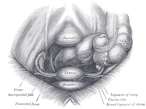 Female pelvis and its contents, seen from above and in frontHenry Vandyke Carter, Gray's Anatomy of the Human Body, 20th ed. (1918), Fig. 1165. SourcePublic domain · Public domain in the US: published before January 1, 1931 (per Wikimedia Commons)Used in: Broad ligament, Ovary
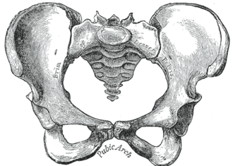 Female pelvis, anterior viewHenry Vandyke Carter, Gray's Anatomy of the Human Body, 20th ed. (1918), Fig. 242. SourcePublic domain · Public domain in the US: published before January 1, 1931 (per Wikimedia Commons)Used in: Bony pelvis
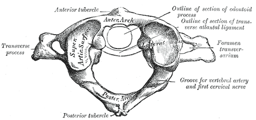 First cervical vertebra, or atlas, from aboveHenry Vandyke Carter, Gray's Anatomy of the Human Body, 20th ed. (1918), Fig. 86. SourcePublic domain · Public domain in the US: published before January 1, 1931 (per Wikimedia Commons)Used in: Atlas and axis (C1 and C2)
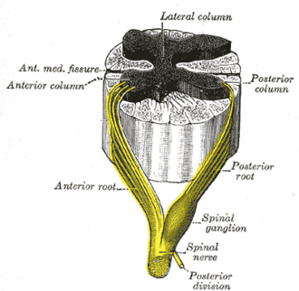 Formation of a spinal nerve from the roots of the spinal cordHenry Vandyke Carter, Gray's Anatomy of the Human Body, 20th ed. (1918), Fig. 675. SourcePublic domain · Public domain in the US: published before January 1, 1931 (per Wikimedia Commons)Used in: Spinal cord, Spinal nerves and roots
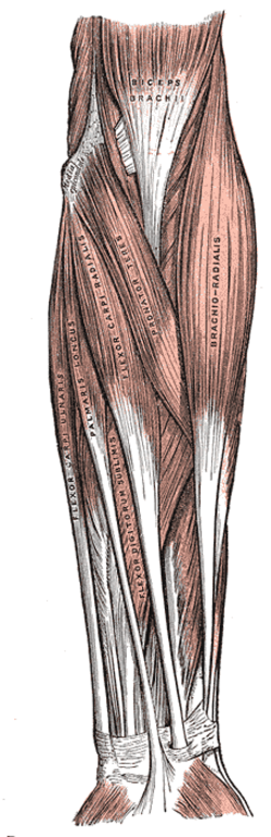 Front of the left forearm, superficial musclesHenry Vandyke Carter, Gray's Anatomy of the Human Body, 20th ed. (1918), Fig. 414. SourcePublic domain · Public domain in the US: published before January 1, 1931 (per Wikimedia Commons)Used in: Pronator teres
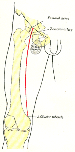 Front of the right thigh: surface markings for the femoral artery and nerveHenry Vandyke Carter, Gray's Anatomy of the Human Body, 20th ed. (1918), Fig. 1245. SourcePublic domain · Public domain in the US: published before January 1, 1931 (per Wikimedia Commons)Used in: Femoral nerve, Femoral triangle
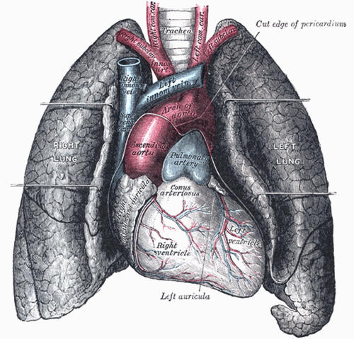 Front view of heart and lungsHenry Vandyke Carter, Gray's Anatomy of the Human Body, 20th ed. (1918), Fig. 970. SourcePublic domain · Public domain in the US: published before January 1, 1931 (per Wikimedia Commons)Used in: Heart, Pericardium
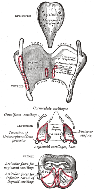
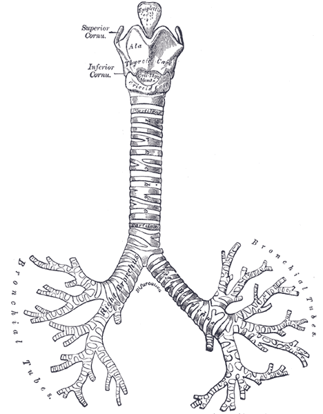 Front view of the cartilages of the larynx, trachea and bronchiHenry Vandyke Carter, Gray's Anatomy of the Human Body, 20th ed. (1918), Fig. 961. SourcePublic domain · Public domain in the US: published before January 1, 1931 (per Wikimedia Commons)Used in: Main bronchi, Trachea
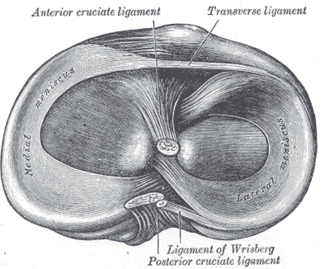 Head of the right tibia from above, showing the menisciHenry Vandyke Carter, Gray's Anatomy of the Human Body, 20th ed. (1918), Fig. 349. SourcePublic domain · Public domain in the US: published before January 1, 1931 (per Wikimedia Commons)Used in: Menisci of the knee
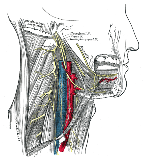 Hypoglossal nerve, cervical plexus and their branchesHenry Vandyke Carter, Gray's Anatomy of the Human Body, 20th ed. (1918), Fig. 794. SourcePublic domain · Public domain in the US: published before January 1, 1931 (per Wikimedia Commons)
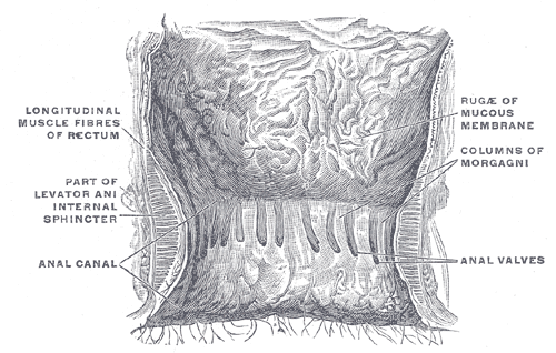 Interior of the anal canal and lower rectumHenry Vandyke Carter, Gray's Anatomy of the Human Body, 20th ed. (1918), Fig. 1080. SourcePublic domain · Public domain in the US: published before January 1, 1931 (per Wikimedia Commons)Used in: Anal canal
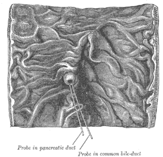 Interior of the descending duodenum showing the bile papillaHenry Vandyke Carter, Gray's Anatomy of the Human Body, 20th ed. (1918), Fig. 1057. SourcePublic domain · Public domain in the US: published before January 1, 1931 (per Wikimedia Commons)Used in: Duodenum, Gallbladder and biliary tree
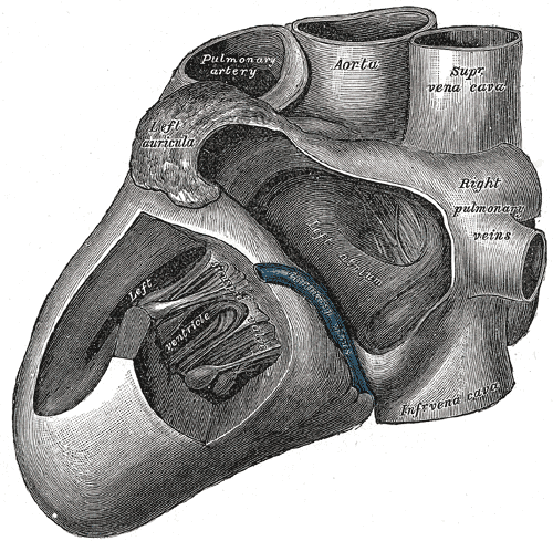 Interior of the left side of the heartHenry Vandyke Carter, Gray's Anatomy of the Human Body, 20th ed. (1918), Fig. 496. SourcePublic domain · Public domain in the US: published before January 1, 1931 (per Wikimedia Commons)Used in: Mitral valve
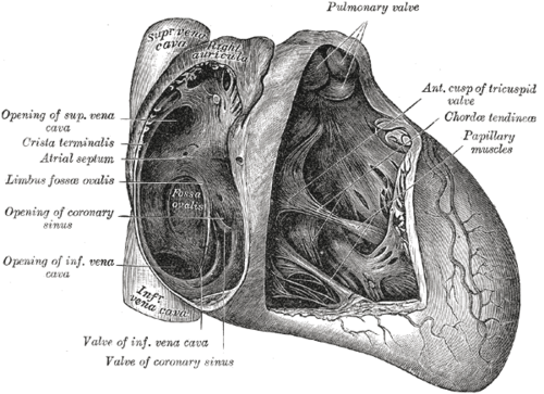 Interior of the right side of the heartHenry Vandyke Carter, Gray's Anatomy of the Human Body, 20th ed. (1918), Fig. 493. SourcePublic domain · Public domain in the US: published before January 1, 1931 (per Wikimedia Commons)Used in: Cardiac conduction system, Tricuspid valve
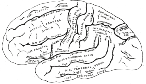 Lateral surface of the left cerebral hemisphereHenry Vandyke Carter, Gray's Anatomy of the Human Body, 20th ed. (1918), Fig. 726. SourcePublic domain · Public domain in the US: published before January 1, 1931 (per Wikimedia Commons)Used in: Cerebral cortex and lobes
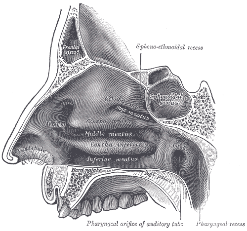 Lateral wall of the nasal cavityHenry Vandyke Carter, Gray's Anatomy of the Human Body, 20th ed. (1918), Fig. 855. SourcePublic domain · Public domain in the US: published before January 1, 1931 (per Wikimedia Commons)Used in: Nasal cavity, Paranasal sinuses
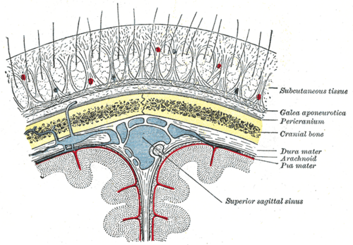 Layers of the scalp, skull and meningesHenry Vandyke Carter, Gray's Anatomy of the Human Body, 20th ed. (1918), Fig. 1196. SourcePublic domain · Public domain in the US: published before January 1, 1931 (per Wikimedia Commons)
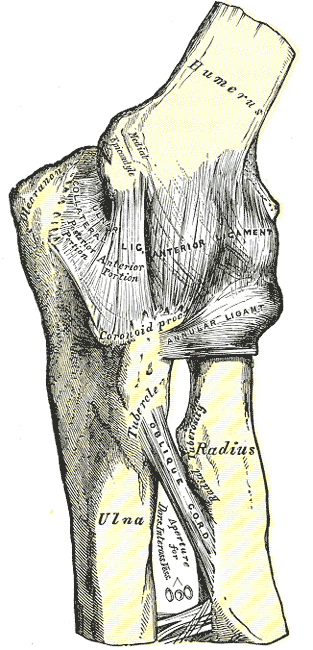 Left elbow joint, anterior and internal (medial) ligamentsHenry Vandyke Carter, Gray's Anatomy of the Human Body, 20th ed. (1918), Fig. 329. SourcePublic domain · Public domain in the US: published before January 1, 1931 (per Wikimedia Commons)Used in: Elbow joint
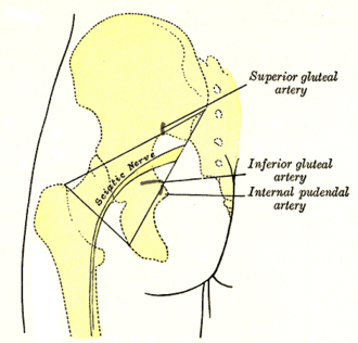 Left gluteal region: surface markings for the arteries and sciatic nerveHenry Vandyke Carter, Gray's Anatomy of the Human Body, 20th ed. (1918), Fig. 1244. SourcePublic domain · Public domain in the US: published before January 1, 1931 (per Wikimedia Commons)Used in: Piriformis, Sciatic nerve
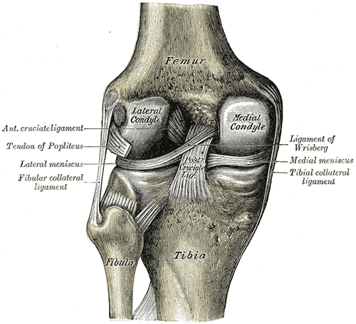 Left knee joint from behind, showing interior ligamentsHenry Vandyke Carter, Gray's Anatomy of the Human Body, 20th ed. (1918), Fig. 348. SourcePublic domain · Public domain in the US: published before January 1, 1931 (per Wikimedia Commons)
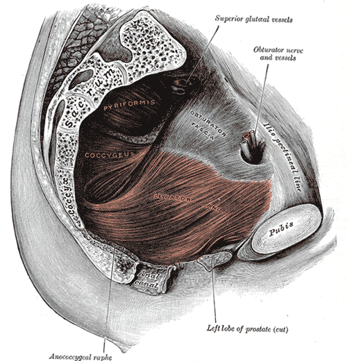 Left levator ani from withinHenry Vandyke Carter, Gray's Anatomy of the Human Body, 20th ed. (1918), Fig. 404. SourcePublic domain · Public domain in the US: published before January 1, 1931 (per Wikimedia Commons)Used in: Pelvic diaphragm
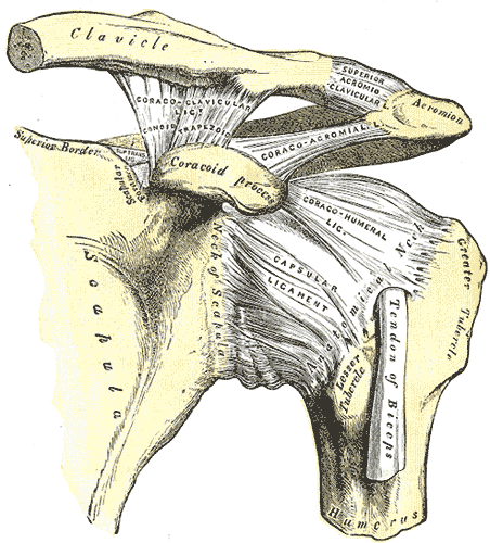 Left shoulder and acromioclavicular joints and the ligaments of the scapulaHenry Vandyke Carter, Gray's Anatomy of the Human Body, 20th ed. (1918), Fig. 326. SourcePublic domain · Public domain in the US: published before January 1, 1931 (per Wikimedia Commons)Used in: Glenohumeral joint
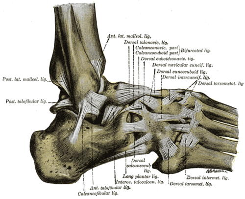 Ligaments of the foot from the lateral aspect (after Quain)Henry Vandyke Carter, Gray's Anatomy of the Human Body, 20th ed. (1918), Fig. 355. SourcePublic domain · Public domain in the US: published before January 1, 1931 (per Wikimedia Commons)Used in: Lateral ankle ligaments
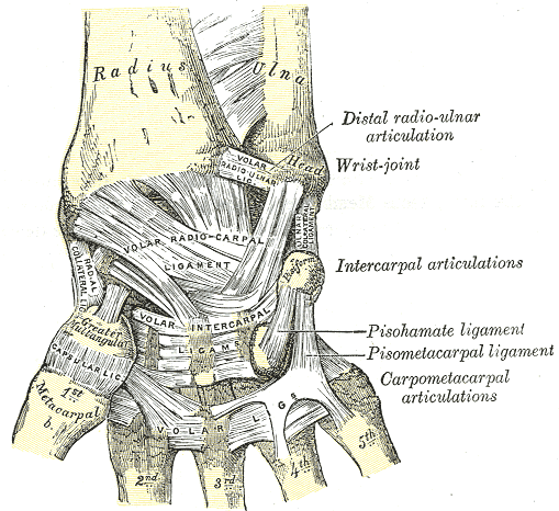 Ligaments of the wrist, anterior viewHenry Vandyke Carter, Gray's Anatomy of the Human Body, 20th ed. (1918), Fig. 334. SourcePublic domain · Public domain in the US: published before January 1, 1931 (per Wikimedia Commons)Used in: Wrist joint
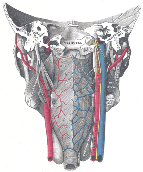
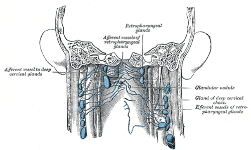 Lymph glands of the pharynx from behindHenry Vandyke Carter, Gray's Anatomy of the Human Body, 20th ed. (1918), Fig. 603. SourcePublic domain · Public domain in the US: published before January 1, 1931 (per Wikimedia Commons)Used in: Cervical lymph nodes, Deep neck spaces
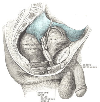 Male pelvic organs seen from the right sideHenry Vandyke Carter, Gray's Anatomy of the Human Body, 20th ed. (1918), Fig. 1136. SourcePublic domain · Public domain in the US: published before January 1, 1931 (per Wikimedia Commons)Used in: Prostate, Urinary bladder
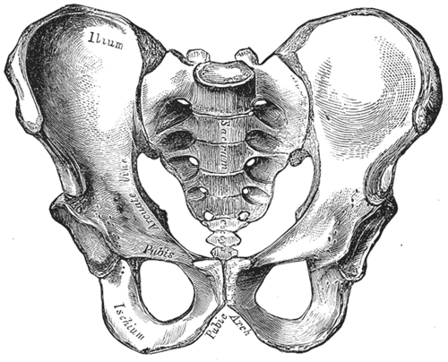 Male pelvis, anterior viewHenry Vandyke Carter, Gray's Anatomy of the Human Body, 20th ed. (1918), Fig. 241. SourcePublic domain · Public domain in the US: published before January 1, 1931 (per Wikimedia Commons)Used in: Bony pelvis
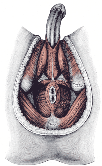
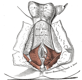 Male perineum: superficial fascia reflectedHenry Vandyke Carter, Gray's Anatomy of the Human Body, 20th ed. (1918), Fig. 405. SourcePublic domain · Public domain in the US: published before January 1, 1931 (per Wikimedia Commons)Used in: Ischioanal fossa, Perineal pouches
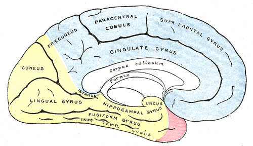 Medial surface of the cerebral hemisphere showing areas supplied by the cerebral arteriesHenry Vandyke Carter, Gray's Anatomy of the Human Body, 20th ed. (1918), Fig. 518. SourcePublic domain · Public domain in the US: published before January 1, 1931 (per Wikimedia Commons)Used in: Cerebral arteries (ACA, MCA, PCA)
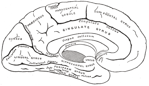 Medial surface of the left cerebral hemisphereHenry Vandyke Carter, Gray's Anatomy of the Human Body, 20th ed. (1918), Fig. 727. SourcePublic domain · Public domain in the US: published before January 1, 1931 (per Wikimedia Commons)Used in: Cerebral cortex and lobes
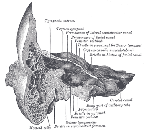 Medial wall of the tympanic cavityHenry Vandyke Carter, Gray's Anatomy of the Human Body, 20th ed. (1918), Fig. 913. SourcePublic domain · Public domain in the US: published before January 1, 1931 (per Wikimedia Commons)Used in: External and middle ear
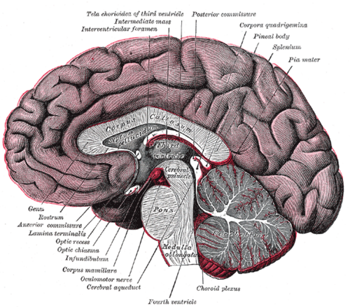 Median sagittal section of the brainHenry Vandyke Carter, Gray's Anatomy of the Human Body, 20th ed. (1918), Fig. 720. SourcePublic domain · Public domain in the US: published before January 1, 1931 (per Wikimedia Commons)
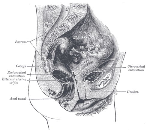 Median sagittal section of the female pelvisHenry Vandyke Carter, Gray's Anatomy of the Human Body, 20th ed. (1918), Fig. 1139. SourcePublic domain · Public domain in the US: published before January 1, 1931 (per Wikimedia Commons)Used in: Rectouterine pouch, Vagina
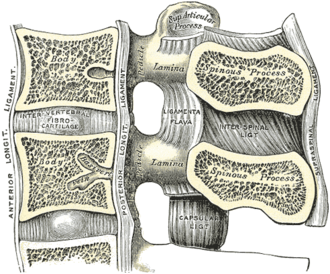 Median sagittal section of two lumbar vertebrae and their ligamentsHenry Vandyke Carter, Gray's Anatomy of the Human Body, 20th ed. (1918), Fig. 301. SourcePublic domain · Public domain in the US: published before January 1, 1931 (per Wikimedia Commons)
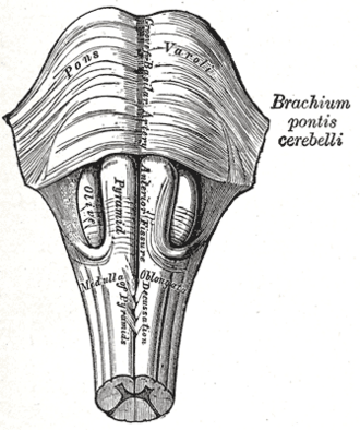 Medulla oblongata and pons, anterior surfaceHenry Vandyke Carter, Gray's Anatomy of the Human Body, 20th ed. (1918), Fig. 679. SourcePublic domain · Public domain in the US: published before January 1, 1931 (per Wikimedia Commons)Used in: Medulla oblongata, Pons
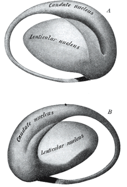 Model of the striatumHenry Vandyke Carter, Gray's Anatomy of the Human Body, 20th ed. (1918), Fig. 741. SourcePublic domain · Public domain in the US: published before January 1, 1931 (per Wikimedia Commons)Used in: Basal ganglia
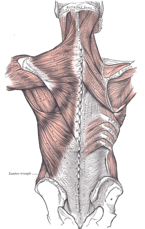 Muscles connecting the upper extremity to the vertebral columnHenry Vandyke Carter, Gray's Anatomy of the Human Body, 20th ed. (1918), Fig. 409. SourcePublic domain · Public domain in the US: published before January 1, 1931 (per Wikimedia Commons)
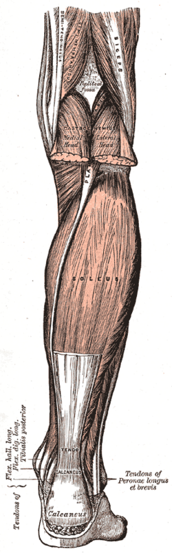 Muscles of the back of the leg, superficial layerHenry Vandyke Carter, Gray's Anatomy of the Human Body, 20th ed. (1918), Fig. 438. SourcePublic domain · Public domain in the US: published before January 1, 1931 (per Wikimedia Commons)Used in: Triceps surae and calcaneal tendon
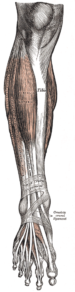 Muscles of the front of the legHenry Vandyke Carter, Gray's Anatomy of the Human Body, 20th ed. (1918), Fig. 437. SourcePublic domain · Public domain in the US: published before January 1, 1931 (per Wikimedia Commons)Used in: Compartments of the leg
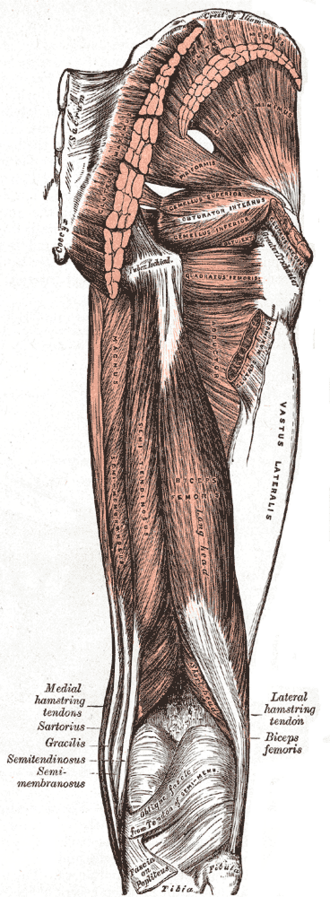 Muscles of the gluteal and posterior femoral regionsHenry Vandyke Carter, Gray's Anatomy of the Human Body, 20th ed. (1918), Fig. 434. SourcePublic domain · Public domain in the US: published before January 1, 1931 (per Wikimedia Commons)Used in: Gluteus maximus, Hamstrings
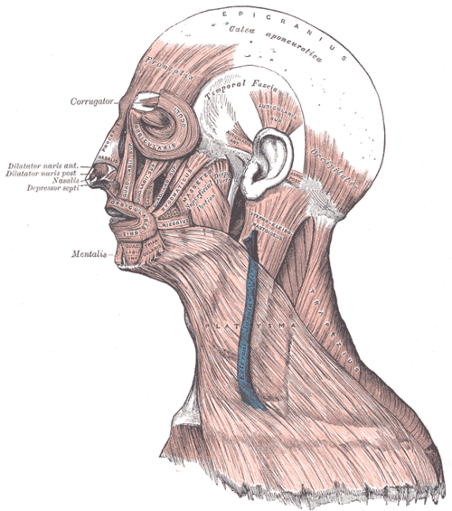 Muscles of the head, face and neckHenry Vandyke Carter, Gray's Anatomy of the Human Body, 20th ed. (1918), Fig. 378. SourcePublic domain · Public domain in the US: published before January 1, 1931 (per Wikimedia Commons)Used in: Muscles of facial expression
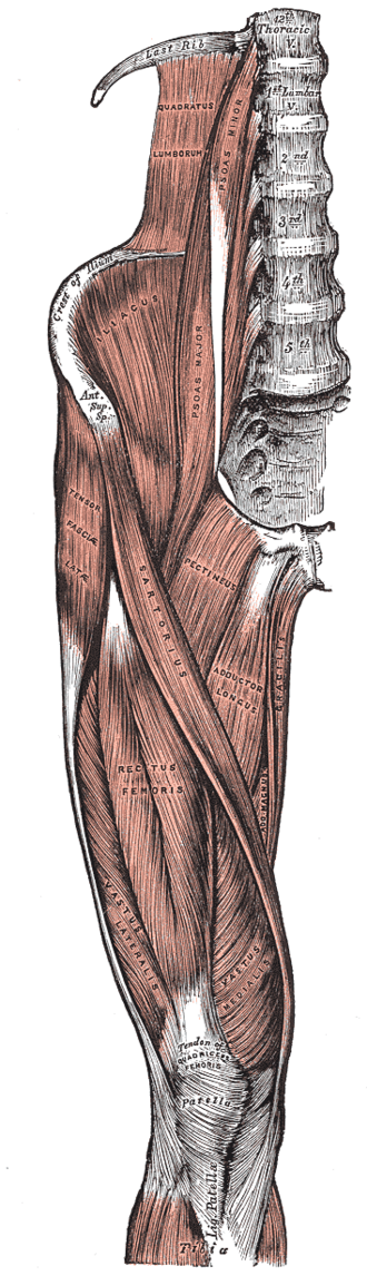 Muscles of the iliac and anterior femoral regionsHenry Vandyke Carter, Gray's Anatomy of the Human Body, 20th ed. (1918), Fig. 430. SourcePublic domain · Public domain in the US: published before January 1, 1931 (per Wikimedia Commons)
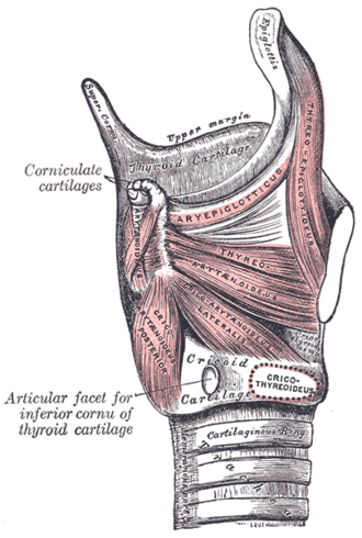 Muscles of the larynx, side viewHenry Vandyke Carter, Gray's Anatomy of the Human Body, 20th ed. (1918), Fig. 959. SourcePublic domain · Public domain in the US: published before January 1, 1931 (per Wikimedia Commons)Used in: Larynx, Recurrent laryngeal nerve
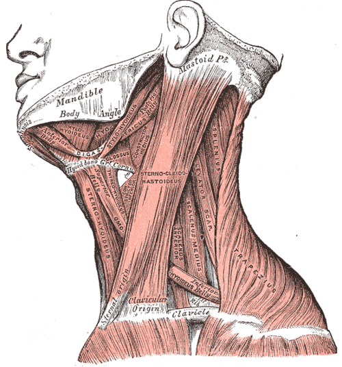 Muscles of the neck, lateral viewHenry Vandyke Carter, Gray's Anatomy of the Human Body, 20th ed. (1918), Fig. 385. SourcePublic domain · Public domain in the US: published before January 1, 1931 (per Wikimedia Commons)Used in: Sternocleidomastoid, Triangles of the neck
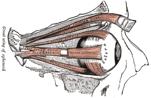 Muscles of the right orbit, lateral viewHenry Vandyke Carter, Gray's Anatomy of the Human Body, 20th ed. (1918), Fig. 889. SourcePublic domain · Public domain in the US: published before January 1, 1931 (per Wikimedia Commons)Used in: Extraocular muscles
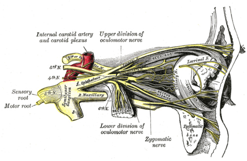 Nerves of the orbit and the ciliary ganglion, side viewHenry Vandyke Carter, Gray's Anatomy of the Human Body, 20th ed. (1918), Fig. 777. SourcePublic domain · Public domain in the US: published before January 1, 1931 (per Wikimedia Commons)
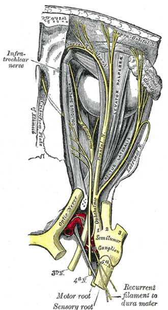 Nerves of the orbit seen from aboveHenry Vandyke Carter, Gray's Anatomy of the Human Body, 20th ed. (1918), Fig. 776. SourcePublic domain · Public domain in the US: published before January 1, 1931 (per Wikimedia Commons)
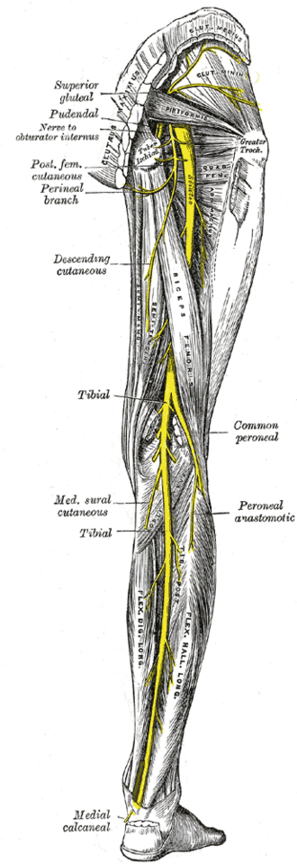 Nerves of the right lower limb, posterior viewHenry Vandyke Carter, Gray's Anatomy of the Human Body, 20th ed. (1918), Fig. 832. SourcePublic domain · Public domain in the US: published before January 1, 1931 (per Wikimedia Commons)Used in: Sciatic nerve, Tibial nerve
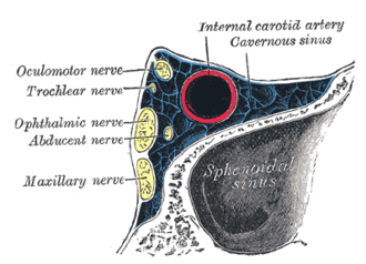 Oblique section through the right cavernous sinusHenry Vandyke Carter, Gray's Anatomy of the Human Body, 20th ed. (1918), Fig. 786. SourcePublic domain · Public domain in the US: published before January 1, 1931 (per Wikimedia Commons)
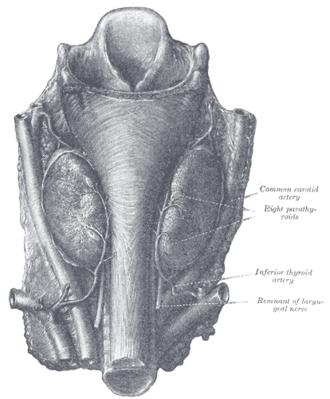 Parathyroid glands (Halsted and Evans)Henry Vandyke Carter, Gray's Anatomy of the Human Body, 20th ed. (1918), Fig. 1177. SourcePublic domain · Public domain in the US: published before January 1, 1931 (per Wikimedia Commons)Used in: Parathyroid glands
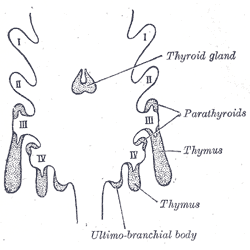 Pharyngeal pouches and their derivativesHenry Vandyke Carter, Gray's Anatomy of the Human Body, 20th ed. (1918), Fig. 1175. SourcePublic domain · Public domain in the US: published before January 1, 1931 (per Wikimedia Commons)Used in: Parathyroid glands
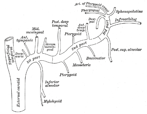 Plan of the branches of the maxillary arteryHenry Vandyke Carter, Gray's Anatomy of the Human Body, 20th ed. (1918), Fig. 511. SourcePublic domain · Public domain in the US: published before January 1, 1931 (per Wikimedia Commons)
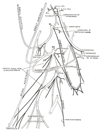 Plan of the cervical plexus (Gerrish)Henry Vandyke Carter, Gray's Anatomy of the Human Body, 20th ed. (1918), Fig. 804. SourcePublic domain · Public domain in the US: published before January 1, 1931 (per Wikimedia Commons)Used in: Cervical plexus
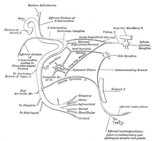 Plan of the facial and intermediate nervesHenry Vandyke Carter, Gray's Anatomy of the Human Body, 20th ed. (1918), Fig. 788. SourcePublic domain · Public domain in the US: published before January 1, 1931 (per Wikimedia Commons)Used in: Facial nerve (CN VII)
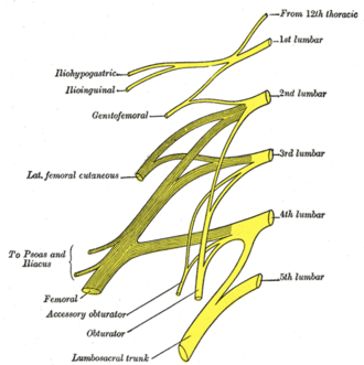 Plan of the lumbar plexusHenry Vandyke Carter, Gray's Anatomy of the Human Body, 20th ed. (1918), Fig. 822. SourcePublic domain · Public domain in the US: published before January 1, 1931 (per Wikimedia Commons)Used in: Lumbar plexus
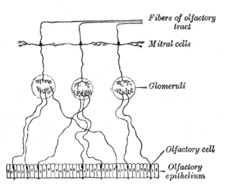 Plan of the olfactory neuronsHenry Vandyke Carter, Gray's Anatomy of the Human Body, 20th ed. (1918), Fig. 772. SourcePublic domain · Public domain in the US: published before January 1, 1931 (per Wikimedia Commons)Used in: Olfactory nerve (CN I)
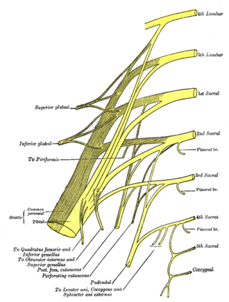 Plan of the sacral and pudendal plexusesHenry Vandyke Carter, Gray's Anatomy of the Human Body, 20th ed. (1918), Fig. 828. SourcePublic domain · Public domain in the US: published before January 1, 1931 (per Wikimedia Commons)Used in: Sacral plexus
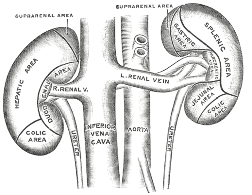
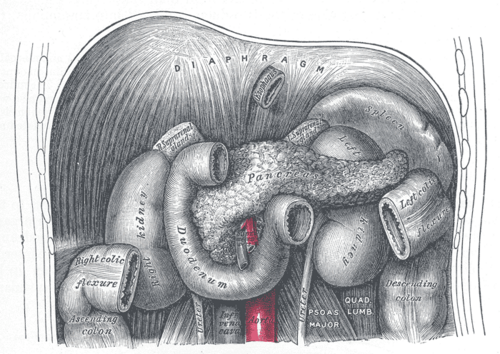
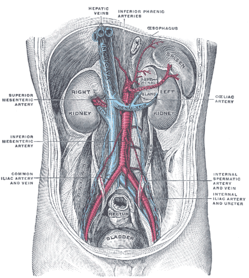 Posterior abdominal wall showing kidneys, suprarenal glands and great vesselsHenry Vandyke Carter, Gray's Anatomy of the Human Body, 20th ed. (1918), Fig. 1121. SourcePublic domain · Public domain in the US: published before January 1, 1931 (per Wikimedia Commons)
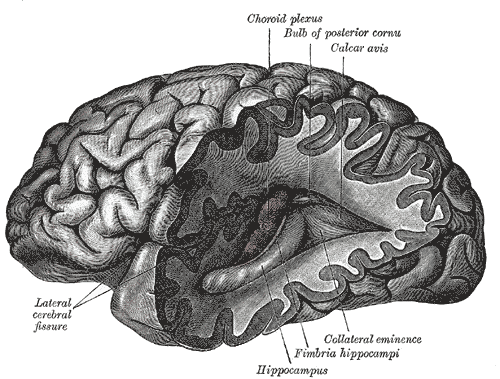 Posterior and inferior horns of the left lateral ventricleHenry Vandyke Carter, Gray's Anatomy of the Human Body, 20th ed. (1918), Fig. 739. SourcePublic domain · Public domain in the US: published before January 1, 1931 (per Wikimedia Commons)Used in: Limbic system
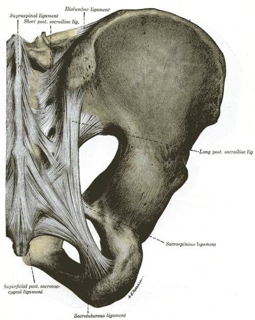 Posterior articulations of the pelvis (after Quain)Henry Vandyke Carter, Gray's Anatomy of the Human Body, 20th ed. (1918), Fig. 320. SourcePublic domain · Public domain in the US: published before January 1, 1931 (per Wikimedia Commons)
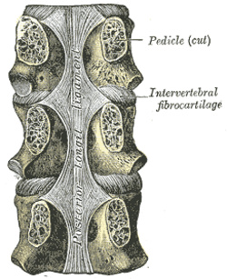 Posterior longitudinal ligament, in the thoracic regionHenry Vandyke Carter, Gray's Anatomy of the Human Body, 20th ed. (1918), Fig. 302. SourcePublic domain · Public domain in the US: published before January 1, 1931 (per Wikimedia Commons)Used in: Vertebral ligaments
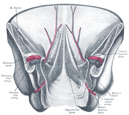 Posterior view of the lower anterior abdominal wallHenry Vandyke Carter, Gray's Anatomy of the Human Body, 20th ed. (1918), Fig. 1036. SourcePublic domain · Public domain in the US: published before January 1, 1931 (per Wikimedia Commons)
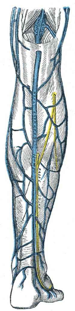 Posterior view of the right leg: small saphenous vein and sural nerveHenry Vandyke Carter, Gray's Anatomy of the Human Body, 20th ed. (1918), Fig. 582. SourcePublic domain · Public domain in the US: published before January 1, 1931 (per Wikimedia Commons)Used in: Deep veins of the lower limb
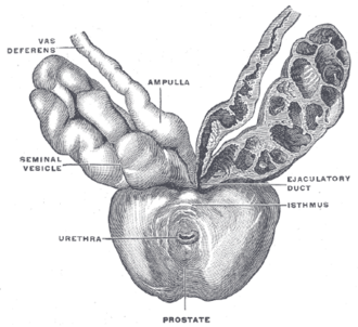 Prostate with seminal vesicles and seminal ducts (after Spalteholz)Henry Vandyke Carter, Gray's Anatomy of the Human Body, 20th ed. (1918), Fig. 1160. SourcePublic domain · Public domain in the US: published before January 1, 1931 (per Wikimedia Commons)Used in: Ductus deferens and seminal glands, Prostate
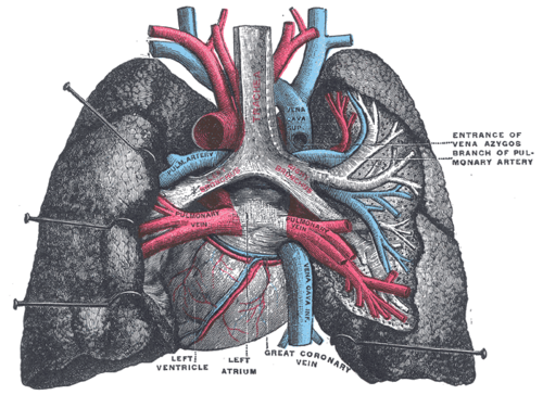 Pulmonary vessels, dorsal view of the heart and lungsHenry Vandyke Carter, Gray's Anatomy of the Human Body, 20th ed. (1918), Fig. 971. SourcePublic domain · Public domain in the US: published before January 1, 1931 (per Wikimedia Commons)Used in: Pulmonary arteries
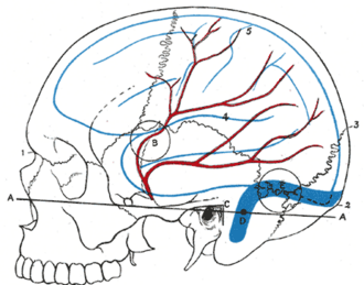 Relations of the brain and middle meningeal artery to the surface of the skullHenry Vandyke Carter, Gray's Anatomy of the Human Body, 20th ed. (1918), Fig. 1198. SourcePublic domain · Public domain in the US: published before January 1, 1931 (per Wikimedia Commons)
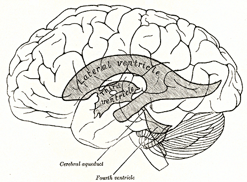 Relations of the ventricles to the surface of the brainHenry Vandyke Carter, Gray's Anatomy of the Human Body, 20th ed. (1918), Fig. 734. SourcePublic domain · Public domain in the US: published before January 1, 1931 (per Wikimedia Commons)Used in: Ventricular system and CSF
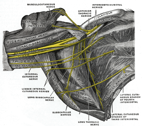 Right brachial plexus (infraclavicular part) in the axilla, from below and in frontHenry Vandyke Carter, Gray's Anatomy of the Human Body, 20th ed. (1918), Fig. 809. SourcePublic domain · Public domain in the US: published before January 1, 1931 (per Wikimedia Commons)
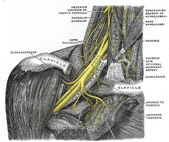 Right brachial plexus with its short branches, anterior viewHenry Vandyke Carter, Gray's Anatomy of the Human Body, 20th ed. (1918), Fig. 808. SourcePublic domain · Public domain in the US: published before January 1, 1931 (per Wikimedia Commons)Used in: Brachial plexus
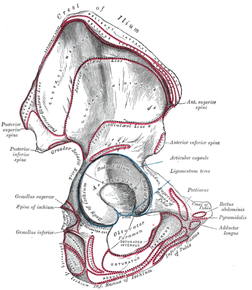
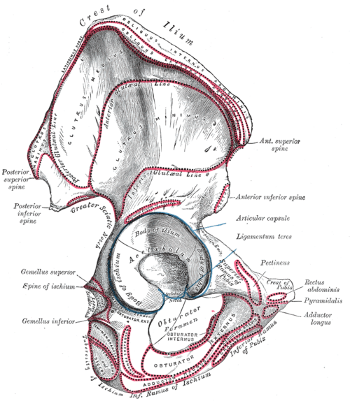 Right hip bone, external surface, with muscle attachmentsHenry Vandyke Carter, Gray's Anatomy of the Human Body, 20th ed. (1918), Fig. 235. SourcePublic domain · Public domain in the US: published before January 1, 1931 (per Wikimedia Commons)Used in: Gluteus medius and minimus
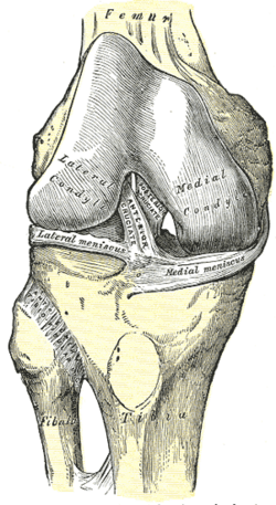 Right knee joint from the front, showing interior ligamentsHenry Vandyke Carter, Gray's Anatomy of the Human Body, 20th ed. (1918), Fig. 347. SourcePublic domain · Public domain in the US: published before January 1, 1931 (per Wikimedia Commons)Used in: Cruciate ligaments, Menisci of the knee
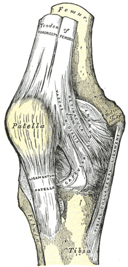 Right knee joint, anterior viewHenry Vandyke Carter, Gray's Anatomy of the Human Body, 20th ed. (1918), Fig. 345. SourcePublic domain · Public domain in the US: published before January 1, 1931 (per Wikimedia Commons)Used in: Collateral ligaments of the knee, Knee joint
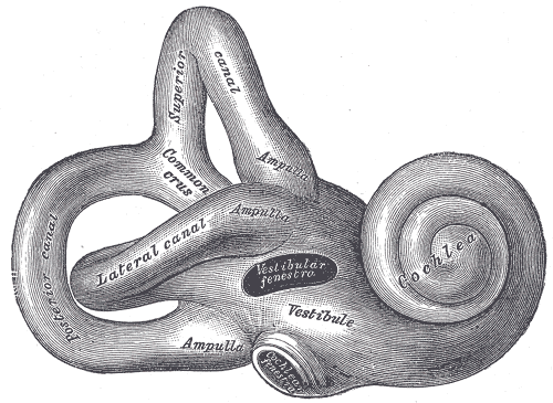 Right osseous labyrinth, lateral viewHenry Vandyke Carter, Gray's Anatomy of the Human Body, 20th ed. (1918), Fig. 920. SourcePublic domain · Public domain in the US: published before January 1, 1931 (per Wikimedia Commons)Used in: Inner ear, Vestibulocochlear nerve (CN VIII)
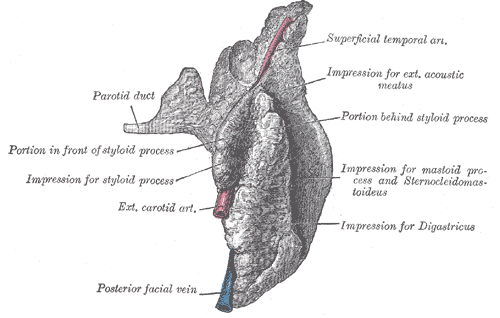 Right parotid gland, posterior and deep aspectsHenry Vandyke Carter, Gray's Anatomy of the Human Body, 20th ed. (1918), Fig. 1022. SourcePublic domain · Public domain in the US: published before January 1, 1931 (per Wikimedia Commons)Used in: Parotid gland
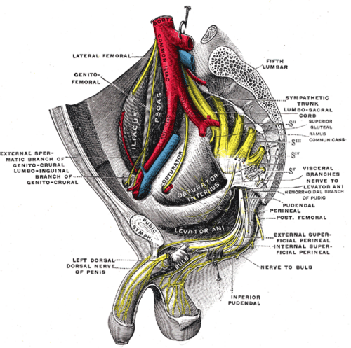 Sacral plexus of the right side (after Testut)Henry Vandyke Carter, Gray's Anatomy of the Human Body, 20th ed. (1918), Fig. 837. SourcePublic domain · Public domain in the US: published before January 1, 1931 (per Wikimedia Commons)Used in: Pudendal nerve
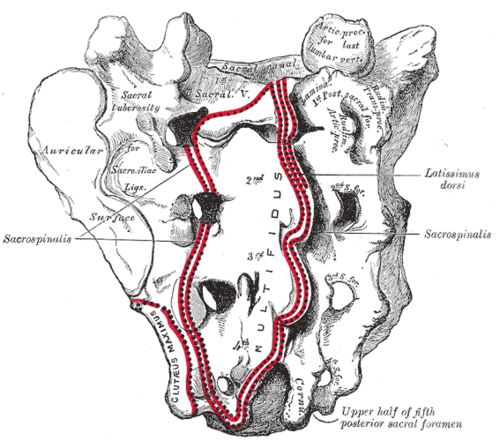 Sacrum, dorsal surfaceHenry Vandyke Carter, Gray's Anatomy of the Human Body, 20th ed. (1918), Fig. 96. SourcePublic domain · Public domain in the US: published before January 1, 1931 (per Wikimedia Commons)Used in: Sacrum and coccyx
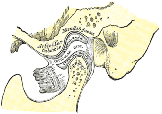 Sagittal section of the articulation of the mandibleHenry Vandyke Carter, Gray's Anatomy of the Human Body, 20th ed. (1918), Fig. 311. SourcePublic domain · Public domain in the US: published before January 1, 1931 (per Wikimedia Commons)Used in: Temporomandibular joint
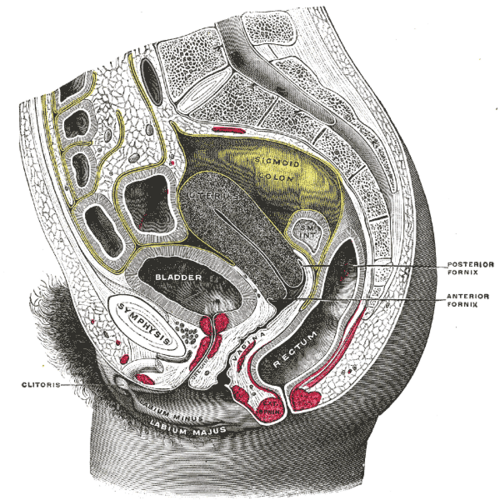 Sagittal section of the lower female trunk, right segment (after Testut)Henry Vandyke Carter, Gray's Anatomy of the Human Body, 20th ed. (1918), Fig. 1166. SourcePublic domain · Public domain in the US: published before January 1, 1931 (per Wikimedia Commons)
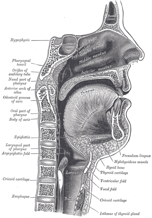 Sagittal section of the nose, mouth, pharynx and larynxHenry Vandyke Carter, Gray's Anatomy of the Human Body, 20th ed. (1918), Fig. 994. SourcePublic domain · Public domain in the US: published before January 1, 1931 (per Wikimedia Commons)
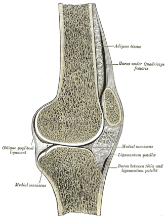 Sagittal section of the right knee jointHenry Vandyke Carter, Gray's Anatomy of the Human Body, 20th ed. (1918), Fig. 350. SourcePublic domain · Public domain in the US: published before January 1, 1931 (per Wikimedia Commons)Used in: Knee joint, Patella
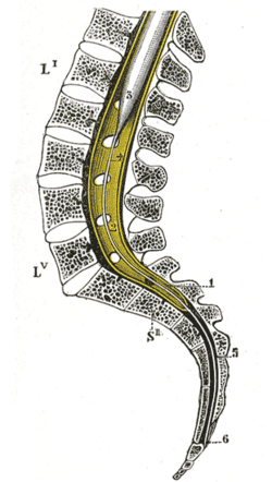 Sagittal section of the vertebral canal showing the lower end of the spinal cord and the filum terminaleHenry Vandyke Carter, Gray's Anatomy of the Human Body, 20th ed. (1918), Fig. 661. SourcePublic domain · Public domain in the US: published before January 1, 1931 (per Wikimedia Commons)Used in: Cauda equina, Spinal meninges and spaces
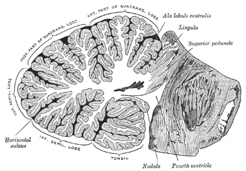 Sagittal section through the cerebellumHenry Vandyke Carter, Gray's Anatomy of the Human Body, 20th ed. (1918), Fig. 704. SourcePublic domain · Public domain in the US: published before January 1, 1931 (per Wikimedia Commons)Used in: Cerebellum
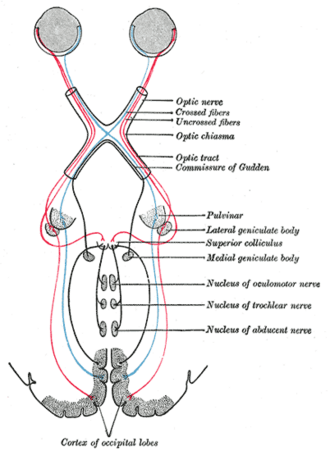 Scheme of the central connections of the optic nerves and tractsHenry Vandyke Carter, Gray's Anatomy of the Human Body, 20th ed. (1918), Fig. 774. SourcePublic domain · Public domain in the US: published before January 1, 1931 (per Wikimedia Commons)
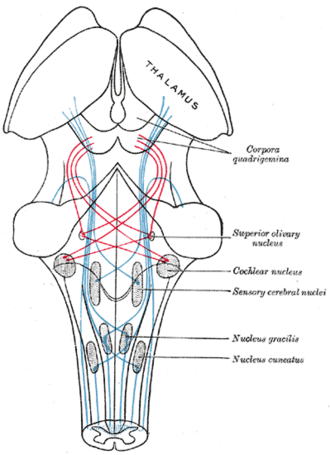 Scheme of the lemniscusHenry Vandyke Carter, Gray's Anatomy of the Human Body, 20th ed. (1918), Fig. 713. SourcePublic domain · Public domain in the US: published before January 1, 1931 (per Wikimedia Commons)
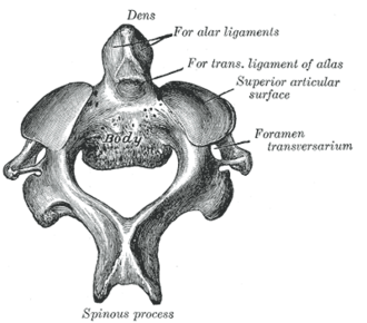 Second cervical vertebra (axis), from aboveHenry Vandyke Carter, Gray's Anatomy of the Human Body, 20th ed. (1918), Fig. 87. SourcePublic domain · Public domain in the US: published before January 1, 1931 (per Wikimedia Commons)Used in: Atlas and axis (C1 and C2)
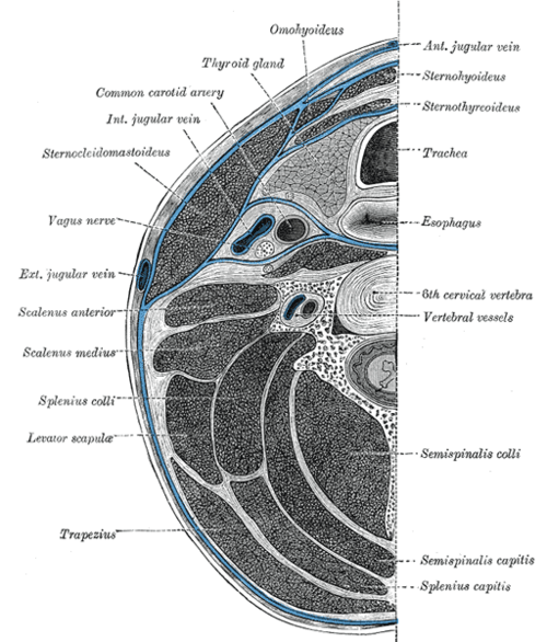 Section of the neck at about the level of the sixth cervical vertebraHenry Vandyke Carter, Gray's Anatomy of the Human Body, 20th ed. (1918), Fig. 384. SourcePublic domain · Public domain in the US: published before January 1, 1931 (per Wikimedia Commons)
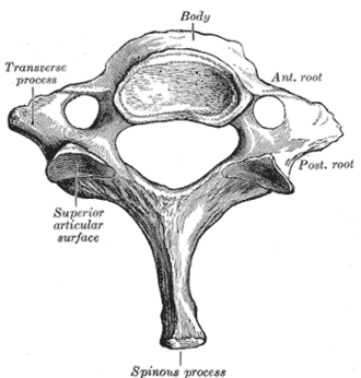 Seventh cervical vertebra (vertebra prominens)Henry Vandyke Carter, Gray's Anatomy of the Human Body, 20th ed. (1918), Fig. 89. SourcePublic domain · Public domain in the US: published before January 1, 1931 (per Wikimedia Commons)Used in: Cervical vertebrae
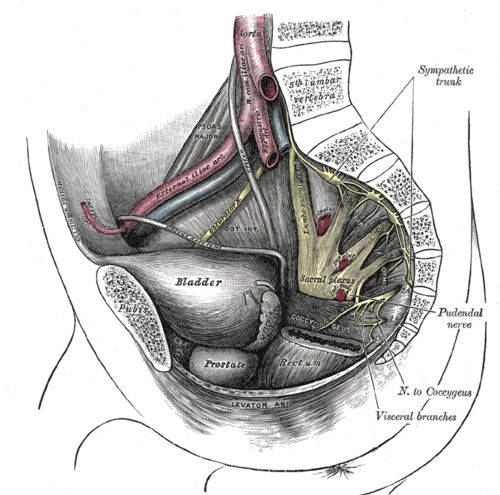 Side wall of the pelvis showing the sacral and pudendal plexuses (after Testut)Henry Vandyke Carter, Gray's Anatomy of the Human Body, 20th ed. (1918), Fig. 829. SourcePublic domain · Public domain in the US: published before January 1, 1931 (per Wikimedia Commons)
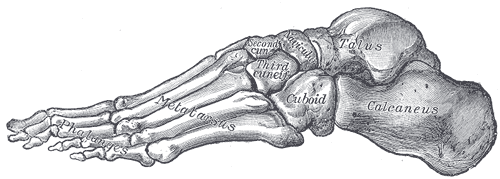 Skeleton of the foot, lateral aspectHenry Vandyke Carter, Gray's Anatomy of the Human Body, 20th ed. (1918), Fig. 291. SourcePublic domain · Public domain in the US: published before January 1, 1931 (per Wikimedia Commons)Used in: Bones of the foot
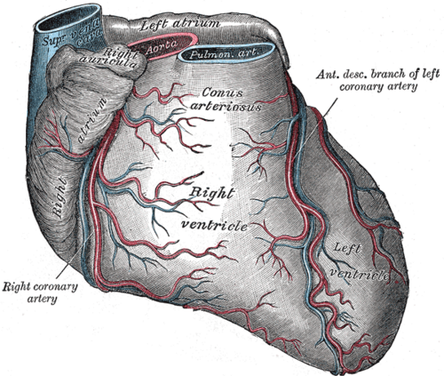 Sternocostal surface of the heart with the coronary arteriesHenry Vandyke Carter, Gray's Anatomy of the Human Body, 20th ed. (1918), Fig. 492. SourcePublic domain · Public domain in the US: published before January 1, 1931 (per Wikimedia Commons)
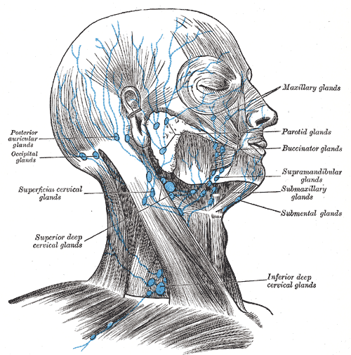 Superficial lymph glands and vessels of the head and neckHenry Vandyke Carter, Gray's Anatomy of the Human Body, 20th ed. (1918), Fig. 602. SourcePublic domain · Public domain in the US: published before January 1, 1931 (per Wikimedia Commons)Used in: Cervical lymph nodes
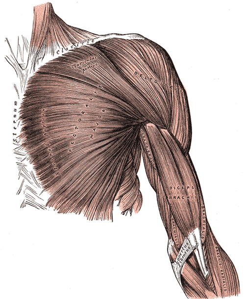 Superficial muscles of the chest and front of the armHenry Vandyke Carter, Gray's Anatomy of the Human Body, 20th ed. (1918), Fig. 410. SourcePublic domain · Public domain in the US: published before January 1, 1931 (per Wikimedia Commons)
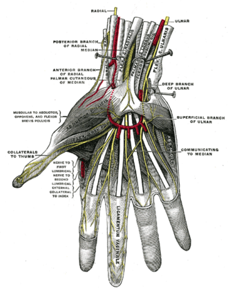 Superficial palmar nerves (Testut)Henry Vandyke Carter, Gray's Anatomy of the Human Body, 20th ed. (1918), Fig. 815. SourcePublic domain · Public domain in the US: published before January 1, 1931 (per Wikimedia Commons)Used in: Median nerve
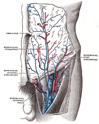 Superficial veins near the saphenofemoral junctionHenry Vandyke Carter, Gray's Anatomy of the Human Body, 20th ed. (1918), Fig. 584. SourcePublic domain · Public domain in the US: published before January 1, 1931 (per Wikimedia Commons)Used in: Great saphenous vein
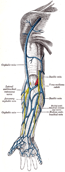 Superficial veins of the upper limbHenry Vandyke Carter, Gray's Anatomy of the Human Body, 20th ed. (1918), Fig. 574. SourcePublic domain · Public domain in the US: published before January 1, 1931 (per Wikimedia Commons)Used in: Cubital fossa
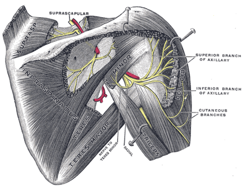 Suprascapular and axillary nerves, from behind (Testut)Henry Vandyke Carter, Gray's Anatomy of the Human Body, 20th ed. (1918), Fig. 810. SourcePublic domain · Public domain in the US: published before January 1, 1931 (per Wikimedia Commons)Used in: Axillary nerve, Deltoid
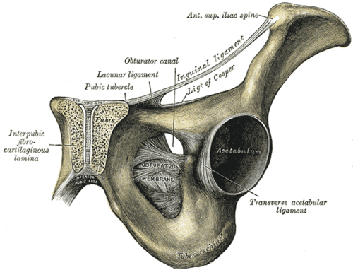 Symphysis pubis exposed by a coronal sectionHenry Vandyke Carter, Gray's Anatomy of the Human Body, 20th ed. (1918), Fig. 321. SourcePublic domain · Public domain in the US: published before January 1, 1931 (per Wikimedia Commons)Used in: Pubic symphysis
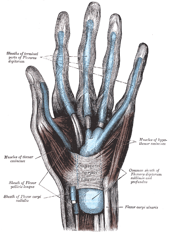 Synovial sheaths of the tendons on the front of the wrist and digitsHenry Vandyke Carter, Gray's Anatomy of the Human Body, 20th ed. (1918), Fig. 1233. SourcePublic domain · Public domain in the US: published before January 1, 1931 (per Wikimedia Commons)Used in: Carpal tunnel
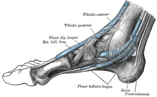 Tendon sheaths around the ankle, medial aspectHenry Vandyke Carter, Gray's Anatomy of the Human Body, 20th ed. (1918), Fig. 442. SourcePublic domain · Public domain in the US: published before January 1, 1931 (per Wikimedia Commons)Used in: Ankle joint
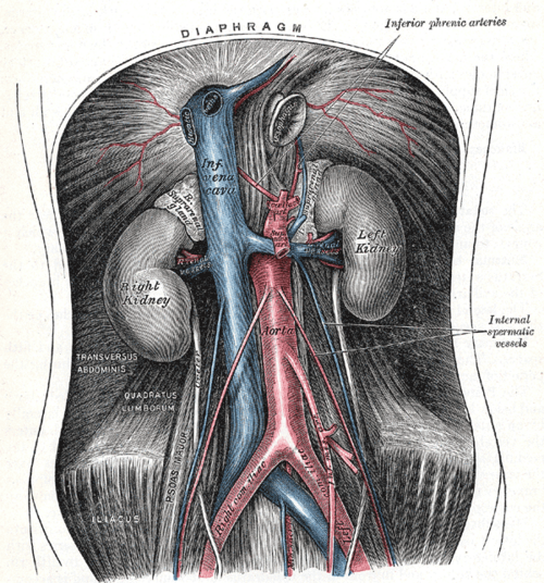 The abdominal aorta and its branchesHenry Vandyke Carter, Gray's Anatomy of the Human Body, 20th ed. (1918), Fig. 531. SourcePublic domain · Public domain in the US: published before January 1, 1931 (per Wikimedia Commons)Used in: Abdominal aorta, Inferior vena cava
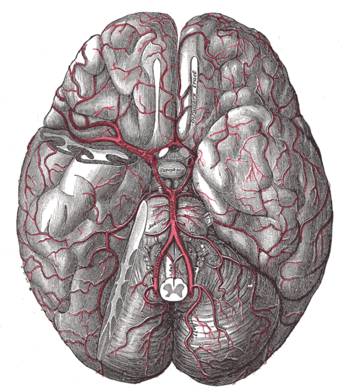 The arteries of the base of the brainHenry Vandyke Carter, Gray's Anatomy of the Human Body, 20th ed. (1918), Fig. 516. SourcePublic domain · Public domain in the US: published before January 1, 1931 (per Wikimedia Commons)
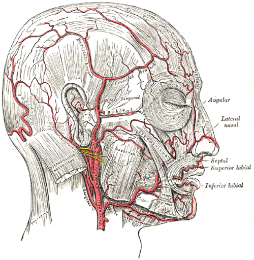 The arteries of the face and scalpHenry Vandyke Carter, Gray's Anatomy of the Human Body, 20th ed. (1918), Fig. 508. SourcePublic domain · Public domain in the US: published before January 1, 1931 (per Wikimedia Commons)Used in: External carotid artery, Scalp
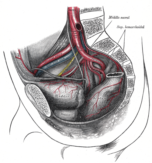 The arteries of the pelvis (male)Henry Vandyke Carter, Gray's Anatomy of the Human Body, 20th ed. (1918), Fig. 539. SourcePublic domain · Public domain in the US: published before January 1, 1931 (per Wikimedia Commons)
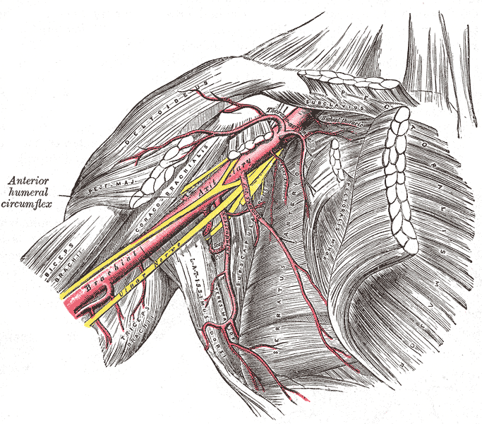 The axillary artery and its branchesHenry Vandyke Carter, Gray's Anatomy of the Human Body, 20th ed. (1918), Fig. 523. SourcePublic domain · Public domain in the US: published before January 1, 1931 (per Wikimedia Commons)Used in: Axillary artery
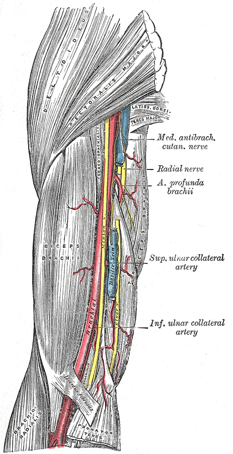 The brachial artery in the arm, medial viewHenry Vandyke Carter, Gray's Anatomy of the Human Body, 20th ed. (1918), Fig. 525. SourcePublic domain · Public domain in the US: published before January 1, 1931 (per Wikimedia Commons)
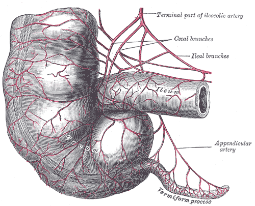 The cecum and vermiform process with their arteriesHenry Vandyke Carter, Gray's Anatomy of the Human Body, 20th ed. (1918), Fig. 1073. SourcePublic domain · Public domain in the US: published before January 1, 1931 (per Wikimedia Commons)Used in: Colon, Vermiform appendix
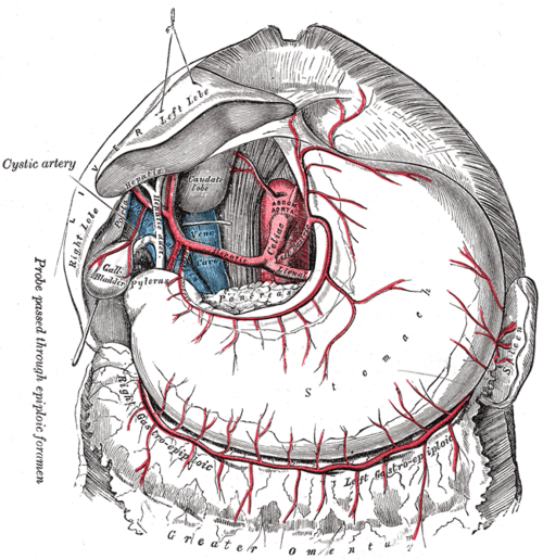 The celiac artery and its branchesHenry Vandyke Carter, Gray's Anatomy of the Human Body, 20th ed. (1918), Fig. 532. SourcePublic domain · Public domain in the US: published before January 1, 1931 (per Wikimedia Commons)
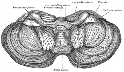 The cerebellum, anterior aspectHenry Vandyke Carter, Gray's Anatomy of the Human Body, 20th ed. (1918), Fig. 703. SourcePublic domain · Public domain in the US: published before January 1, 1931 (per Wikimedia Commons)Used in: Cerebellum
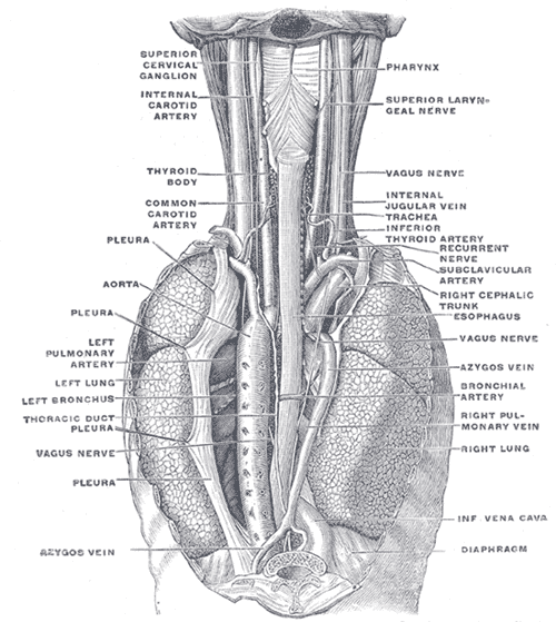 The esophagus in the neck and posterior mediastinum, from behindHenry Vandyke Carter, Gray's Anatomy of the Human Body, 20th ed. (1918), Fig. 1032. SourcePublic domain · Public domain in the US: published before January 1, 1931 (per Wikimedia Commons)Used in: Esophagus, Thoracic duct
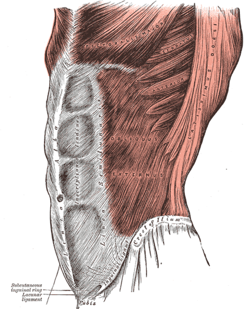 The external oblique and the rectus sheathHenry Vandyke Carter, Gray's Anatomy of the Human Body, 20th ed. (1918), Fig. 392. SourcePublic domain · Public domain in the US: published before January 1, 1931 (per Wikimedia Commons)Used in: Rectus abdominis
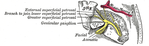 The facial nerve in the temporal boneHenry Vandyke Carter, Gray's Anatomy of the Human Body, 20th ed. (1918), Fig. 789. SourcePublic domain · Public domain in the US: published before January 1, 1931 (per Wikimedia Commons)Used in: Facial nerve (CN VII)
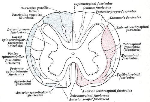 The fasciculi of the spinal cordHenry Vandyke Carter, Gray's Anatomy of the Human Body, 20th ed. (1918), Fig. 672. SourcePublic domain · Public domain in the US: published before January 1, 1931 (per Wikimedia Commons)
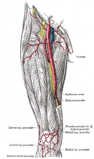 The femoral artery in the thighHenry Vandyke Carter, Gray's Anatomy of the Human Body, 20th ed. (1918), Fig. 550. SourcePublic domain · Public domain in the US: published before January 1, 1931 (per Wikimedia Commons)Used in: Femoral artery
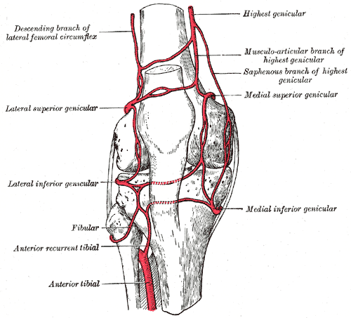 The genicular anastomosis around the kneeHenry Vandyke Carter, Gray's Anatomy of the Human Body, 20th ed. (1918), Fig. 552. SourcePublic domain · Public domain in the US: published before January 1, 1931 (per Wikimedia Commons)Used in: Knee joint
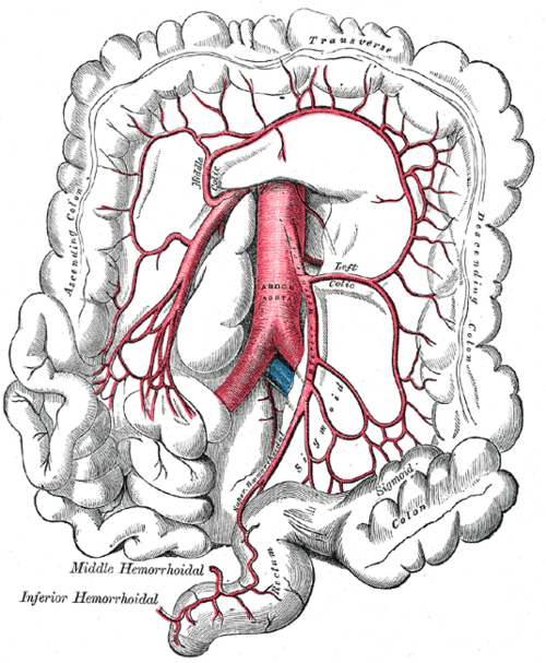 The inferior mesenteric artery and its branchesHenry Vandyke Carter, Gray's Anatomy of the Human Body, 20th ed. (1918), Fig. 537. SourcePublic domain · Public domain in the US: published before January 1, 1931 (per Wikimedia Commons)Used in: Colon, Inferior mesenteric artery
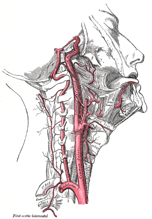 The internal carotid and vertebral arteries, right sideHenry Vandyke Carter, Gray's Anatomy of the Human Body, 20th ed. (1918), Fig. 513. SourcePublic domain · Public domain in the US: published before January 1, 1931 (per Wikimedia Commons)Used in: Vertebral artery
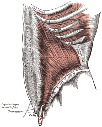 The internal oblique muscleHenry Vandyke Carter, Gray's Anatomy of the Human Body, 20th ed. (1918), Fig. 395. SourcePublic domain · Public domain in the US: published before January 1, 1931 (per Wikimedia Commons)Used in: Rectus abdominis
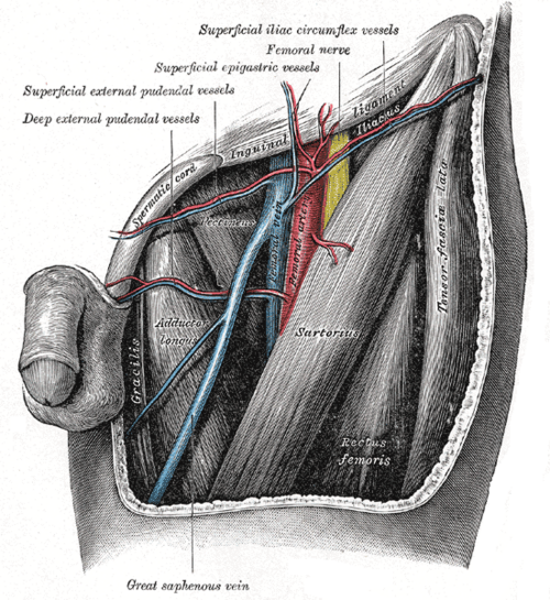 The left femoral triangleHenry Vandyke Carter, Gray's Anatomy of the Human Body, 20th ed. (1918), Fig. 549. SourcePublic domain · Public domain in the US: published before January 1, 1931 (per Wikimedia Commons)Used in: Femoral triangle, Great saphenous vein
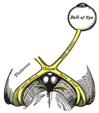 The left optic nerve and the optic tractsHenry Vandyke Carter, Gray's Anatomy of the Human Body, 20th ed. (1918), Fig. 773. SourcePublic domain · Public domain in the US: published before January 1, 1931 (per Wikimedia Commons)
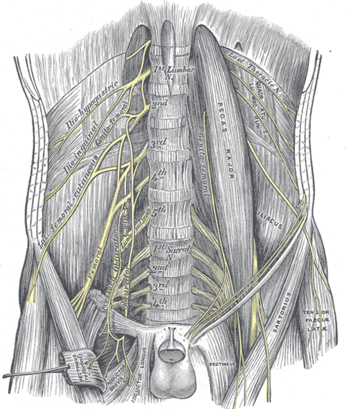 The lumbar plexus and its branchesHenry Vandyke Carter, Gray's Anatomy of the Human Body, 20th ed. (1918), Fig. 823. SourcePublic domain · Public domain in the US: published before January 1, 1931 (per Wikimedia Commons)Used in: Femoral nerve, Obturator nerve
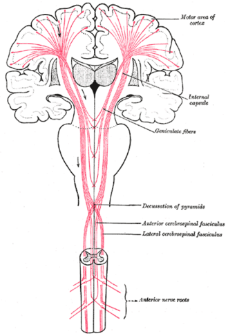 The motor tract (after Poirier)Henry Vandyke Carter, Gray's Anatomy of the Human Body, 20th ed. (1918), Fig. 764. SourcePublic domain · Public domain in the US: published before January 1, 1931 (per Wikimedia Commons)Used in: Corticospinal tract
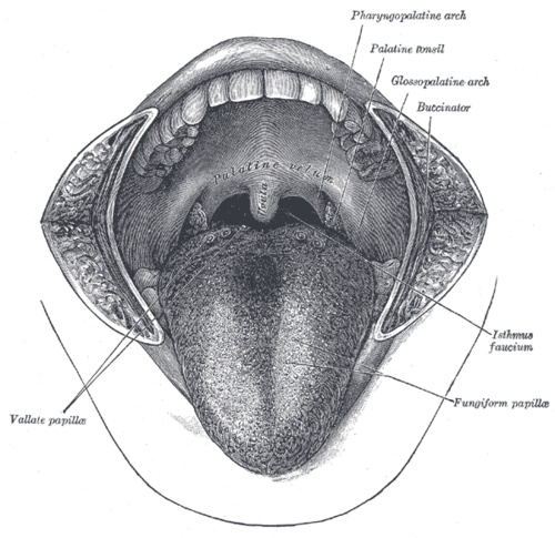 The mouth cavityHenry Vandyke Carter, Gray's Anatomy of the Human Body, 20th ed. (1918), Fig. 1014. SourcePublic domain · Public domain in the US: published before January 1, 1931 (per Wikimedia Commons)Used in: Palatine tonsils, Tongue
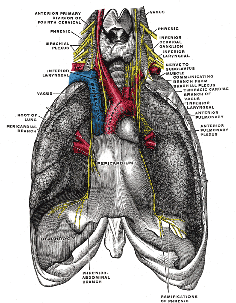 The phrenic nerve and its relations with the vagus nerveHenry Vandyke Carter, Gray's Anatomy of the Human Body, 20th ed. (1918), Fig. 806. SourcePublic domain · Public domain in the US: published before January 1, 1931 (per Wikimedia Commons)Used in: Phrenic nerve
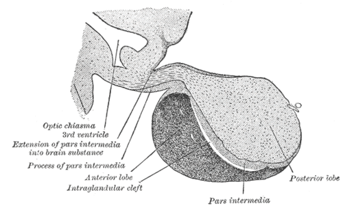 The pituitary gland (median sagittal section)Henry Vandyke Carter, Gray's Anatomy of the Human Body, 20th ed. (1918), Fig. 1181. SourcePublic domain · Public domain in the US: published before January 1, 1931 (per Wikimedia Commons)Used in: Hypothalamus, Pituitary gland
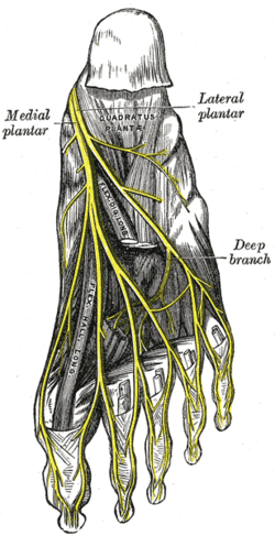 The plantar nervesHenry Vandyke Carter, Gray's Anatomy of the Human Body, 20th ed. (1918), Fig. 833. SourcePublic domain · Public domain in the US: published before January 1, 1931 (per Wikimedia Commons)Used in: Tibial nerve
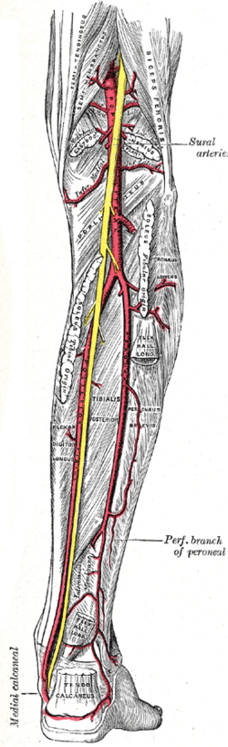 The popliteal and posterior tibial arteriesHenry Vandyke Carter, Gray's Anatomy of the Human Body, 20th ed. (1918), Fig. 551. SourcePublic domain · Public domain in the US: published before January 1, 1931 (per Wikimedia Commons)Used in: Popliteal artery, Popliteal fossa
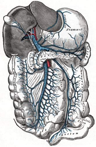 The portal vein and its tributariesHenry Vandyke Carter, Gray's Anatomy of the Human Body, 20th ed. (1918), Fig. 591. SourcePublic domain · Public domain in the US: published before January 1, 1931 (per Wikimedia Commons)Used in: Hepatic portal vein
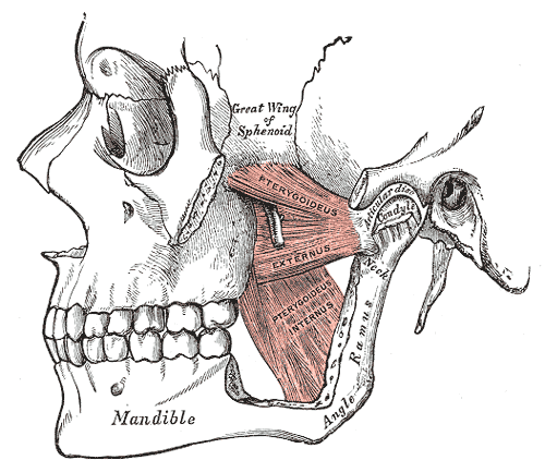 The pterygoid musclesHenry Vandyke Carter, Gray's Anatomy of the Human Body, 20th ed. (1918), Fig. 383. SourcePublic domain · Public domain in the US: published before January 1, 1931 (per Wikimedia Commons)Used in: Muscles of mastication
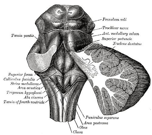 The rhomboid fossa (floor of the fourth ventricle)Henry Vandyke Carter, Gray's Anatomy of the Human Body, 20th ed. (1918), Fig. 709. SourcePublic domain · Public domain in the US: published before January 1, 1931 (per Wikimedia Commons)
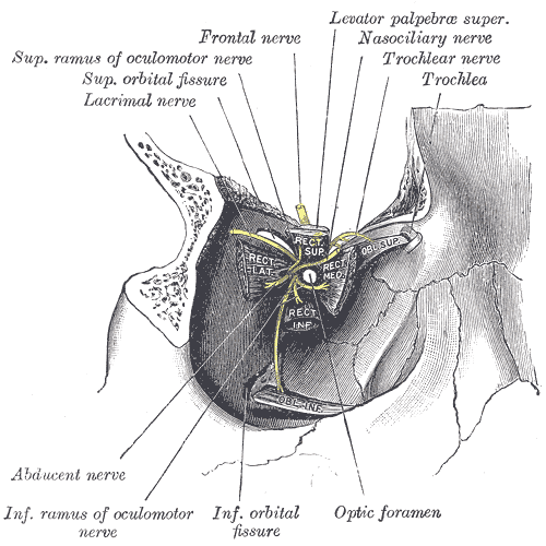 The right orbit from the front with the eyeball removedHenry Vandyke Carter, Gray's Anatomy of the Human Body, 20th ed. (1918), Fig. 890. SourcePublic domain · Public domain in the US: published before January 1, 1931 (per Wikimedia Commons)Used in: Extraocular muscles, Orbit
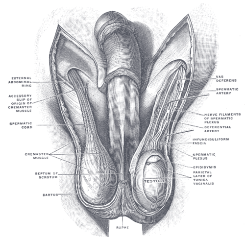 The scrotum and testes, anterior viewUnknown (Gray's Anatomy, 1918; author not recorded on Commons), Gray's Anatomy of the Human Body, 20th ed. (1918), Fig. 1144. SourcePublic domain · Public domain in the US: published before January 1, 1931 (per Wikimedia Commons)Used in: Testis and epididymis
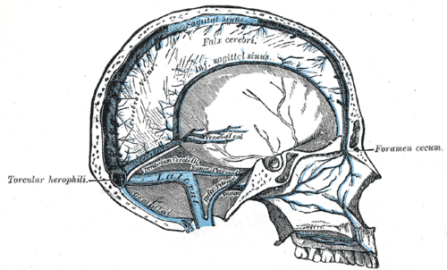 The sinuses of the dura materHenry Vandyke Carter, Gray's Anatomy of the Human Body, 20th ed. (1918), Fig. 568. SourcePublic domain · Public domain in the US: published before January 1, 1931 (per Wikimedia Commons)Used in: Dural venous sinuses
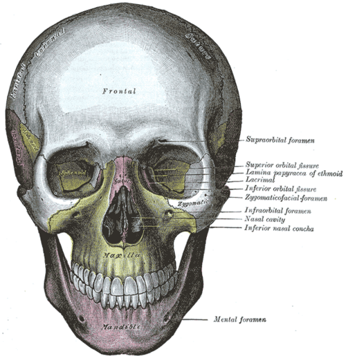 The skull from the frontHenry Vandyke Carter, Gray's Anatomy of the Human Body, 20th ed. (1918), Fig. 190. SourcePublic domain · Public domain in the US: published before January 1, 1931 (per Wikimedia Commons)
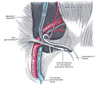 The spermatic cord in the inguinal canalHenry Vandyke Carter, Gray's Anatomy of the Human Body, 20th ed. (1918), Fig. 1146. SourcePublic domain · Public domain in the US: published before January 1, 1931 (per Wikimedia Commons)Used in: Inguinal canal, Spermatic cord
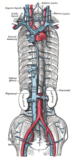 The superior and inferior venae cavae and their tributariesHenry Vandyke Carter, Gray's Anatomy of the Human Body, 20th ed. (1918), Fig. 577. SourcePublic domain · Public domain in the US: published before January 1, 1931 (per Wikimedia Commons)Used in: Azygos vein, Superior vena cava
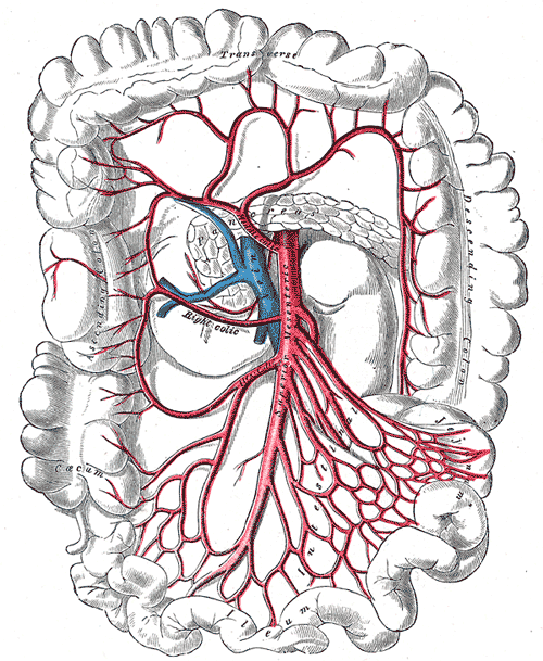 The superior mesenteric artery and its branchesHenry Vandyke Carter, Gray's Anatomy of the Human Body, 20th ed. (1918), Fig. 534. SourcePublic domain · Public domain in the US: published before January 1, 1931 (per Wikimedia Commons)
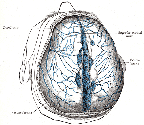 The superior sagittal sinus laid openHenry Vandyke Carter, Gray's Anatomy of the Human Body, 20th ed. (1918), Fig. 566. SourcePublic domain · Public domain in the US: published before January 1, 1931 (per Wikimedia Commons)Used in: Dural venous sinuses
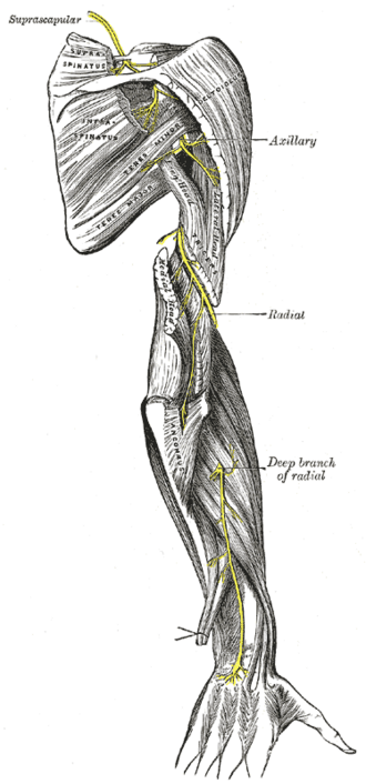 The suprascapular, axillary and radial nervesHenry Vandyke Carter, Gray's Anatomy of the Human Body, 20th ed. (1918), Fig. 818. SourcePublic domain · Public domain in the US: published before January 1, 1931 (per Wikimedia Commons)Used in: Axillary nerve
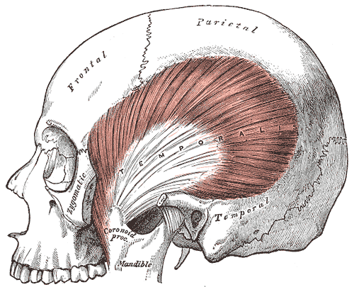 The temporalis muscleHenry Vandyke Carter, Gray's Anatomy of the Human Body, 20th ed. (1918), Fig. 382. SourcePublic domain · Public domain in the US: published before January 1, 1931 (per Wikimedia Commons)Used in: Muscles of mastication
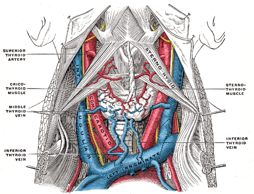 The thyroid gland and its veinsHenry Vandyke Carter, Gray's Anatomy of the Human Body, 20th ed. (1918), Fig. 562. SourcePublic domain · Public domain in the US: published before January 1, 1931 (per Wikimedia Commons)Used in: Internal jugular vein, Thyroid gland
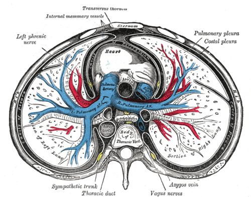 Transverse section of the thoraxHenry Vandyke Carter, Gray's Anatomy of the Human Body, 20th ed. (1918), Fig. 968. SourcePublic domain · Public domain in the US: published before January 1, 1931 (per Wikimedia Commons)Used in: Mediastinum, Pleural cavity
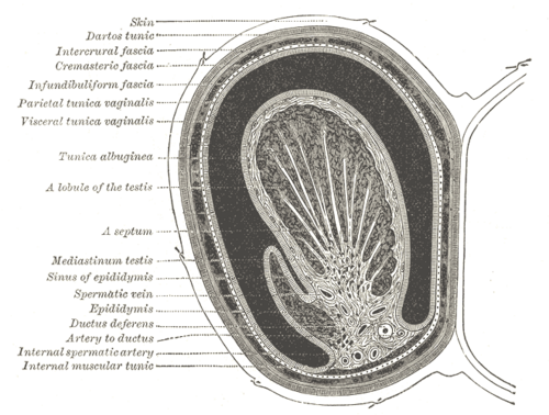 Transverse section through the left scrotum and testisHenry Vandyke Carter, Gray's Anatomy of the Human Body, 20th ed. (1918), Fig. 1145. SourcePublic domain · Public domain in the US: published before January 1, 1931 (per Wikimedia Commons)Used in: Testis and epididymis
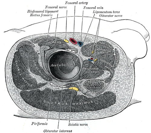 Transverse section through the right hip jointHenry Vandyke Carter, Gray's Anatomy of the Human Body, 20th ed. (1918), Fig. 344. SourcePublic domain · Public domain in the US: published before January 1, 1931 (per Wikimedia Commons)Used in: Gluteus medius and minimus
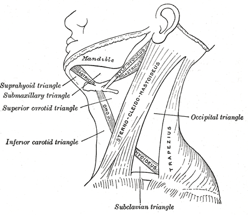 Triangles of the neckHenry Vandyke Carter, Gray's Anatomy of the Human Body, 20th ed. (1918), Fig. 512. SourcePublic domain · Public domain in the US: published before January 1, 1931 (per Wikimedia Commons)Used in: Triangles of the neck
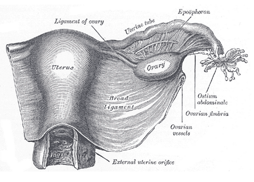 Uterus and right broad ligament, seen from behindHenry Vandyke Carter, Gray's Anatomy of the Human Body, 20th ed. (1918), Fig. 1161. SourcePublic domain · Public domain in the US: published before January 1, 1931 (per Wikimedia Commons)
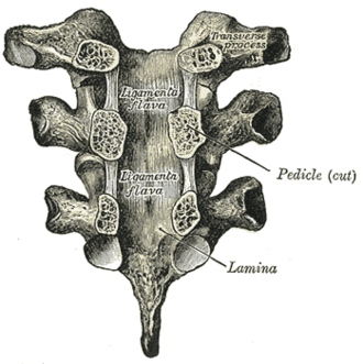 Vertebral arches of three thoracic vertebrae viewed from the frontHenry Vandyke Carter, Gray's Anatomy of the Human Body, 20th ed. (1918), Fig. 303. SourcePublic domain · Public domain in the US: published before January 1, 1931 (per Wikimedia Commons)Used in: Vertebral ligaments
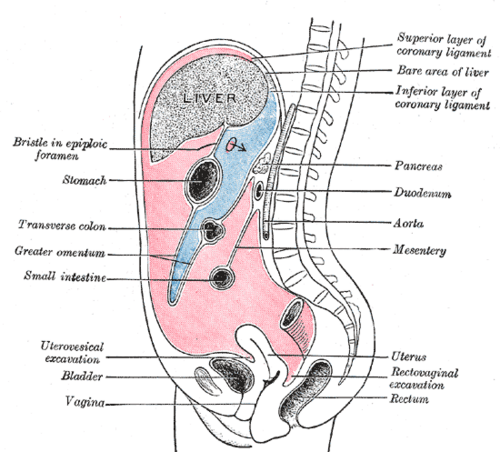 Vertical disposition of the peritoneumHenry Vandyke Carter, Gray's Anatomy of the Human Body, 20th ed. (1918), Fig. 1035. SourcePublic domain · Public domain in the US: published before January 1, 1931 (per Wikimedia Commons)