Limbic system
Network of medial temporal and diencephalic structures for memory, emotion and behavior. The hippocampus and mammillary bodies are damaged in Alzheimer disease, Wernicke–Korsakoff syndrome and HSV encephalitis.
Core anatomy
- Location
Medial rim of the cerebral hemisphere around the diencephalon, mainly the medial temporal lobe.
- Parts
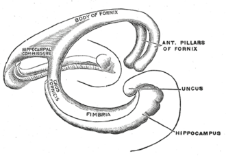
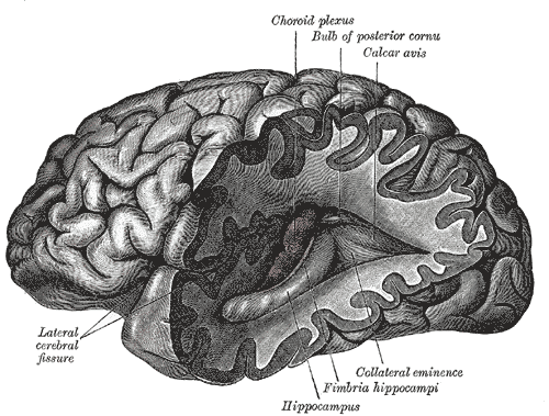
- Hippocampus (medial temporal lobe, floor of the temporal horn) — forms new declarative memories; CA1 neurons are very sensitive to hypoxia
- Amygdala (anterior to the hippocampus) — fear, emotional memory, autonomic responses
- Fornix — output tract of the hippocampus to the mammillary bodies
- Mammillary bodies (hypothalamus) → mammillothalamic tract → anterior nucleus of the thalamus → cingulate gyrus → parahippocampal/entorhinal cortex → hippocampus (Papez circuit)
- Cingulate gyrus, parahippocampal gyrus, septal nuclei, nucleus accumbens (reward), olfactory connections
- Relations
The hippocampus lies in the floor of the temporal horn of the lateral ventricle (see ventricular system and CSF).
- Arterial supply
Posterior cerebral artery (hippocampus), anterior choroidal artery (amygdala, hippocampus), ACA (cingulate) — see cerebral arteries (ACA, MCA, PCA).
- Venous drainage
Basal vein → great cerebral vein.
- Notes
Receives olfactory input directly (olfactory nerve (CN I)); outputs to the hypothalamus and prefrontal cortex.
Mentioned in
Generated automatically from links in other entries.


Clinical correlations
Wernicke encephalopathy and Korsakoff syndrome PANCE
- Mechanism / lesion
Thiamine (B1) deficiency — alcohol use disorder, malnutrition, hyperemesis, bariatric surgery; damages the mammillary bodies and medial thalamus.
- Classic presentation
Wernicke: confusion, ophthalmoplegia/nystagmus, ataxia (not all three are always present) — give IV thiamine before glucose. Korsakoff: permanent anterograde and retrograde amnesia with confabulation.
Alzheimer disease PANCE
- Mechanism / lesion
Amyloid plaques and neurofibrillary tangles; early loss in the hippocampus and entorhinal cortex, cholinergic deficit (nucleus basalis of Meynert).
- Classic presentation
Insidious short-term memory loss first, then language, visuospatial and executive decline; MRI shows hippocampal/medial temporal atrophy. Cholinesterase inhibitors (donepezil), memantine.
HSV encephalitis and temporal lobe epilepsy PANCE
- Mechanism / lesion
HSV-1 has a predilection for the medial temporal and orbitofrontal lobes; mesial temporal sclerosis (hippocampal) is the most common cause of focal epilepsy in adults.
- Classic presentation
HSV encephalitis: fever, headache, confusion, behavior change, seizures, aphasia; MRI temporal lobe changes, CSF HSV PCR — start IV acyclovir immediately. Temporal lobe seizures: aura (déjà vu, rising epigastric sensation, smells), staring, lip-smacking automatisms.
Klüver–Bucy syndrome
- Mechanism / lesion
Bilateral amygdala (anterior temporal) damage, e.g. HSV encephalitis, trauma.
- Classic presentation
Hyperorality, hypersexuality, placidity (loss of fear) and visual agnosia.
High-yield PANCE pearls
- Confusion + ophthalmoplegia + ataxia in an alcoholic → Wernicke → thiamine before glucose.
- Amnesia + confabulation → Korsakoff (mammillary bodies).
- Fever + confusion + temporal lobe changes → HSV encephalitis → IV acyclovir now.
- Déjà vu aura + automatisms → temporal lobe (hippocampal) seizure.
- Early Alzheimer → hippocampal atrophy, short-term memory loss.
Imaging anatomy
- CT
Often normal early in HSV encephalitis and Wernicke encephalopathy.
- MRI
MRI — hippocampal atrophy (Alzheimer), mesial temporal sclerosis (epilepsy protocol), temporal lobe T2/FLAIR signal in HSV encephalitis, mammillary body changes in Wernicke encephalopathy.
- Key views & landmarks
- EEG — temporal spikes; periodic lateralized discharges in HSV encephalitis
Physical exam
- Strength / function testing
Orientation, recall of three words at 5 minutes (anterograde memory), remote memory, cognitive screening (MoCA/MMSE); eye movements and gait (Wernicke); mood and behavior.
Self-quiz
A 50-year-old man with alcohol use disorder is confused, has horizontal nystagmus with bilateral lateral rectus weakness, and a wide-based gait. What is the most appropriate immediate treatment?
Correct answer: B
Wernicke encephalopathy (confusion, ophthalmoplegia, ataxia) from thiamine deficiency; give IV thiamine immediately and before glucose, which can precipitate or worsen it.
Show answer
B
Wernicke encephalopathy (confusion, ophthalmoplegia, ataxia) from thiamine deficiency; give IV thiamine immediately and before glucose, which can precipitate or worsen it.
A 34-year-old has 3 days of fever, headache and odd behavior, then a seizure. MRI shows right medial temporal lobe hyperintensity. What is the most appropriate treatment?
Correct answer: B
HSV-1 encephalitis favors the medial temporal lobes. Start IV acyclovir as soon as it is suspected; confirm with CSF HSV PCR.
Show answer
B
HSV-1 encephalitis favors the medial temporal lobes. Start IV acyclovir as soon as it is suspected; confirm with CSF HSV PCR.
In the Papez circuit, the mammillothalamic tract connects the mammillary bodies to which thalamic nucleus?
Correct answer: C
Papez circuit: hippocampus → fornix → mammillary bodies → mammillothalamic tract → anterior nucleus of the thalamus → cingulate gyrus → parahippocampal cortex → hippocampus.
Show answer
C
Papez circuit: hippocampus → fornix → mammillary bodies → mammillothalamic tract → anterior nucleus of the thalamus → cingulate gyrus → parahippocampal cortex → hippocampus.
After HSV encephalitis, a patient shows hyperorality, hypersexuality, placidity and failure to recognize objects by sight. Which structures are damaged?
Correct answer: B
Klüver–Bucy syndrome results from bilateral amygdala (anterior temporal) damage, e.g. HSV encephalitis or trauma.
Show answer
B
Klüver–Bucy syndrome results from bilateral amygdala (anterior temporal) damage, e.g. HSV encephalitis or trauma.
Which structures show the earliest neuronal loss and atrophy in Alzheimer disease?
Correct answer: D
Alzheimer disease starts in the hippocampus and entorhinal cortex, which is why short-term memory loss comes first; MRI shows medial temporal atrophy.
Show answer
D
Alzheimer disease starts in the hippocampus and entorhinal cortex, which is why short-term memory loss comes first; MRI shows medial temporal atrophy.