Thyroid gland
Butterfly-shaped endocrine gland wrapped around the upper trachea at C5–T1. It moves with swallowing, lies next to the recurrent laryngeal nerves and parathyroids, and thyroid nodules are a common workup.
Core anatomy
- Location
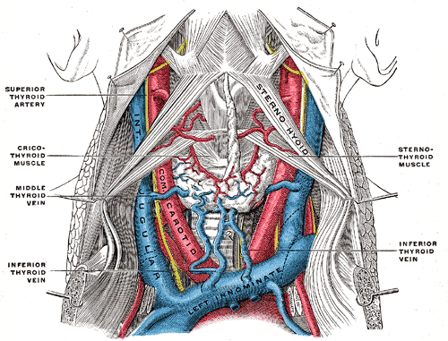
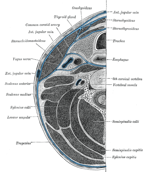
Anterior neck, deep to the infrahyoid (strap) muscles, at about C5–T1; the isthmus lies over the 2nd–3rd (or 2nd–4th) tracheal rings.
- Parts
- Right and left lobes on the sides of the larynx and trachea
- Isthmus over the upper tracheal rings
- Pyramidal lobe (variable) rising from the isthmus — remnant of the thyroglossal duct
- Enclosed in the pretracheal layer of deep cervical fascia, which attaches it to the larynx, so it moves up on swallowing
- Follicular cells (T3/T4) and parafollicular C cells (calcitonin)
- Relations
Posteromedially: the recurrent laryngeal nerve in the tracheoesophageal groove, close to the inferior thyroid artery. Posteriorly: the parathyroid glands. The external laryngeal nerve runs near the superior thyroid vessels. Posterolaterally: the carotid sheath (common and internal carotid arteries, internal jugular vein, vagus).
- Arterial supply
Superior thyroid artery (first branch of the external carotid artery) and inferior thyroid artery (from the thyrocervical trunk of the subclavian); occasionally a thyroid ima artery from the brachiocephalic trunk or arch.
- Venous drainage
Superior and middle thyroid veins → internal jugular vein; inferior thyroid veins → brachiocephalic veins.
- Lymphatic drainage
Prelaryngeal (Delphian), pretracheal and paratracheal (central compartment, level VI) nodes, then deep cervical nodes (see cervical lymph nodes).
- Innervation
Sympathetic fibers from the cervical ganglia (vasomotor); secretion is controlled hormonally by TSH.
- Notes
Develops from the foramen cecum of the tongue and descends along the thyroglossal duct; persistence gives a thyroglossal duct cyst.
Forms a boundary of
Found in
Mentioned in
Generated automatically from links in other entries.
Swipe sideways to see the whole diagram →


Clinical correlations
Thyroid nodule PANCE
- Mechanism / lesion
Very common; most are benign (colloid nodules, adenomas); about 5–10% are malignant (papillary carcinoma most common). verify
- Classic presentation
Workup: TSH first. Low TSH → radioactive iodine uptake scan (a "hot" nodule is rarely malignant). Normal or high TSH → ultrasound → fine-needle aspiration based on size and suspicious features (microcalcifications, hypoechoic, irregular margins, taller-than-wide).
Thyroid cancer PANCE
- Mechanism / lesion
Papillary (most common; radiation exposure; psammoma bodies; lymphatic spread), follicular (hematogenous spread), medullary (C cells; calcitonin; MEN 2), anaplastic (elderly; rapidly fatal).
- Classic presentation
A hard, fixed nodule, cervical lymphadenopathy, hoarseness (nerve invasion), rapid growth.
Thyroglossal duct cyst PANCE
- Mechanism / lesion
Persistent thyroglossal duct remnant.
- Classic presentation
Midline neck mass near the hyoid that moves up with tongue protrusion and swallowing; can become infected. Excise with the middle of the hyoid (Sistrunk procedure) after confirming normal thyroid tissue elsewhere.
Goiter and thyroidectomy complications PANCE
- Mechanism / lesion
Goiter — iodine deficiency, Graves disease, multinodular goiter, Hashimoto thyroiditis. Complications of thyroidectomy — recurrent laryngeal nerve injury, hypoparathyroidism, neck hematoma.
- Classic presentation
Neck hematoma after thyroidectomy can compress the airway — open the wound at the bedside. Hypocalcemia (perioral tingling, Chvostek and Trousseau signs) from parathyroid injury. Hoarseness from RLN injury. A large goiter can cause Pemberton sign (facial plethora on raising the arms).
High-yield PANCE pearls
- Thyroid nodule → TSH first; low → uptake scan; normal/high → ultrasound ± FNA.
- Papillary = most common, psammoma bodies, radiation; medullary = calcitonin, MEN 2.
- Midline mass that moves with tongue protrusion → thyroglossal duct cyst.
- Post-thyroidectomy: hoarseness (RLN), hypocalcemia (parathyroids), hematoma (airway).
Imaging anatomy
- X-ray
Tracheal deviation from a large goiter may be seen on a chest X-ray.
- CT
Retrosternal goiter extent, airway compression, and staging of advanced cancer (avoid iodinated contrast before planned radioiodine therapy when possible).
- Ultrasound
First-line imaging for nodules and goiter; guides FNA.
- Key views & landmarks
- Thyroid ultrasound
- Radioactive iodine (I-123) uptake scan
Physical exam
- Surface landmarks
Find the thyroid cartilage, then the cricoid; the isthmus lies just below the cricoid and the lobes lie lateral to it, behind the sternocleidomastoids.
- Palpation
Examine from behind or in front with the neck slightly flexed; have the patient swallow water — the thyroid rises. Assess size, consistency, nodules, tenderness (subacute thyroiditis) and cervical nodes. Auscultate for a bruit (Graves disease).
- Strength / function testing
Look for signs of hypo- or hyperthyroidism (reflexes, tremor, skin, eyes).
Self-quiz
A 42-year-old woman has a 2-cm thyroid nodule. TSH is normal. What is the most appropriate next step?
Correct answer: B
With a normal TSH, the next step is a thyroid ultrasound to characterize the nodule and decide on FNA. An uptake scan is for a low TSH.
Show answer
B
With a normal TSH, the next step is a thyroid ultrasound to characterize the nodule and decide on FNA. An uptake scan is for a low TSH.
A 7-year-old has a painless midline neck mass at the level of the hyoid that rises when she sticks out her tongue. What is the most likely diagnosis?
Correct answer: B
A midline mass that moves with tongue protrusion is a thyroglossal duct cyst, attached to the hyoid and foramen cecum. A branchial cleft cyst lies laterally, along the anterior border of sternocleidomastoid.
Show answer
B
A midline mass that moves with tongue protrusion is a thyroglossal duct cyst, attached to the hyoid and foramen cecum. A branchial cleft cyst lies laterally, along the anterior border of sternocleidomastoid.
Six hours after thyroidectomy, a patient has an expanding neck swelling, stridor and difficulty breathing. What is the most appropriate immediate action?
Correct answer: C
An expanding post-thyroidectomy hematoma compresses the airway; open the wound immediately (skin and strap sutures) to relieve pressure, then return to the OR. Imaging would delay life-saving treatment.
Show answer
C
An expanding post-thyroidectomy hematoma compresses the airway; open the wound immediately (skin and strap sutures) to relieve pressure, then return to the OR. Imaging would delay life-saving treatment.
A patient with a thyroid nodule has a low TSH. What is the most appropriate next step?
Correct answer: B
With a low TSH, a radioactive iodine uptake scan is next; a "hot" (hyperfunctioning) nodule is rarely malignant. With a normal or high TSH, ultrasound ± FNA is next.
Show answer
B
With a low TSH, a radioactive iodine uptake scan is next; a "hot" (hyperfunctioning) nodule is rarely malignant. With a normal or high TSH, ultrasound ± FNA is next.
Medullary thyroid carcinoma arises from which cells, and which tumor marker is used?
Correct answer: E
Medullary carcinoma arises from parafollicular C cells, secretes calcitonin, and is associated with MEN 2. Papillary carcinoma (most common) arises from follicular cells.
Show answer
E
Medullary carcinoma arises from parafollicular C cells, secretes calcitonin, and is associated with MEN 2. Papillary carcinoma (most common) arises from follicular cells.