Teres minor
Small rotator cuff muscle from the lateral border of the scapula. It laterally rotates the arm and is supplied by the axillary nerve.
Core anatomy
- Location
Along the lateral border of the scapula, inferior to infraspinatus (often partly blended with it).
- Group
Rotator cuff (SITS); scapulohumeral muscles.
- Origin
Middle part of the lateral border of the scapula.
- Insertion
Inferior facet of the greater tubercle of the humerus.
- Innervation
Axillary nerve (C5, C6), via its posterior branch
- Blood supply
Circumflex scapular and posterior circumflex humeral arteries verify
- Action
- Laterally (externally) rotates the arm
- With the other rotator cuff muscles, holds the humeral head in the glenoid cavity
- Relations
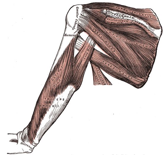
Forms the superior boundary of the quadrangular space (axillary nerve and posterior circumflex humeral artery). Together with teres major and the long head of triceps it also bounds the triangular space (circumflex scapular artery). It is the only rotator cuff muscle not supplied by the suprascapular or subscapular nerves.
Swipe sideways to see the whole diagram →

Clinical correlations
Quadrilateral space syndrome
- Mechanism / lesion
Compression of the axillary nerve in the quadrangular space, often in overhead athletes.
- Classic presentation
Posterior shoulder pain and paresthesias. On MRI, isolated teres minor atrophy/denervation is a characteristic finding.
Axillary nerve injury PANCE
- Mechanism / lesion
Surgical neck fracture of the humerus or anterior shoulder dislocation.
- Classic presentation
Weak lateral rotation (teres minor) together with deltoid weakness and lateral shoulder numbness.
High-yield PANCE pearls
- Teres minor is the only rotator cuff muscle innervated by the axillary nerve.
- Superior border of the quadrangular space.
- Hornblower's sign tests teres minor.
Imaging anatomy
- X-ray
Not assessed directly.
- MRI
Sagittal oblique images show muscle bulk. Isolated fatty atrophy or edema of teres minor suggests axillary nerve compression (quadrilateral space syndrome). Tendon tears are uncommon and usually part of a massive cuff tear.
- Ultrasound
Seen from a posterior approach, inferior to infraspinatus, inserting on the lowest facet of the greater tuberosity.
Physical exam
- Surface landmarks
Lies along the lateral border of the scapula. Difficult to separate from infraspinatus by palpation.
- Strength / function testing
Resisted lateral rotation, especially with the arm abducted (teres minor contributes more in abduction).
Hornblower's sign
- Technique
The arm is supported at 90° of abduction in the scapular plane with the elbow flexed at 90°. The patient tries to externally rotate against resistance (or bring the hand to the mouth).
- Positive test
Unable to externally rotate in this position, or has to abduct the arm further (elbow rising like a horn player's).
- Significance
Teres minor dysfunction, usually with a massive posterosuperior cuff tear.
Self-quiz
Which rotator cuff muscle is innervated by the axillary nerve?
Correct answer: C
Teres minor is supplied by the posterior branch of the axillary nerve. Supraspinatus and infraspinatus are supplied by the suprascapular nerve, subscapularis by the upper and lower subscapular nerves. Teres major is not part of the rotator cuff.
Show answer
C
Teres minor is supplied by the posterior branch of the axillary nerve. Supraspinatus and infraspinatus are supplied by the suprascapular nerve, subscapularis by the upper and lower subscapular nerves. Teres major is not part of the rotator cuff.
Teres minor forms which boundary of the quadrangular space?
Correct answer: A
Quadrangular space boundaries: teres minor (superior), teres major (inferior), long head of triceps (medial), surgical neck of the humerus (lateral).
Show answer
A
Quadrangular space boundaries: teres minor (superior), teres major (inferior), long head of triceps (medial), surgical neck of the humerus (lateral).
MRI of a 24-year-old baseball pitcher with posterior shoulder pain shows fatty atrophy limited to teres minor. Which structure is most likely compressed?
Correct answer: B
Isolated teres minor denervation points to the axillary nerve in the quadrangular space (quadrilateral space syndrome). Deltoid may be involved too.
Show answer
B
Isolated teres minor denervation points to the axillary nerve in the quadrangular space (quadrilateral space syndrome). Deltoid may be involved too.