Rectouterine pouch
Deepest recess of the female peritoneal cavity, between the uterus and rectum. Blood, pus and ascites collect here; the male equivalent is the rectovesical pouch.
Core anatomy
- Location
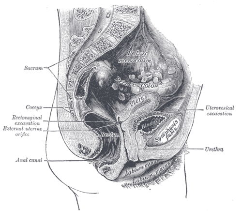
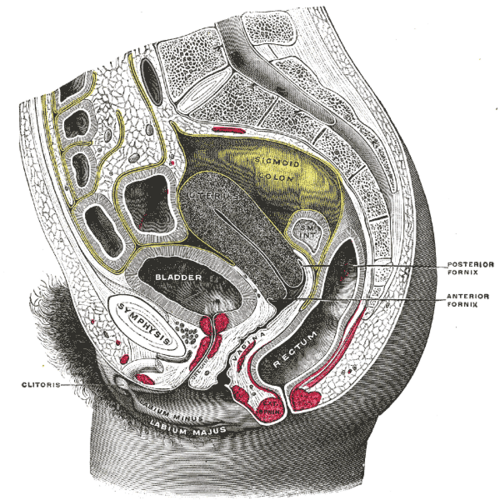
The lowest part of the peritoneal cavity in a woman who is upright or supine, behind the uterus and the posterior fornix of the vagina, in front of the rectum.
- Boundaries
- anterior
- Posterior surface of the uterus, supravaginal cervix and posterior vaginal fornix
- posterior
- Anterior surface of the rectum (middle third)
- lateral
- Rectouterine (uterosacral) folds of peritoneum
- floor
- Peritoneum reflected from the posterior vaginal fornix onto the rectum, about 5–6 cm above the anus verify
- Contents
- Normally loops of small intestine or sigmoid colon and a small amount of physiologic peritoneal fluid
- Pathologically: blood (ruptured ectopic pregnancy or cyst), pus (pelvic abscess, PID), ascites, endometriosis, and peritoneal metastases
- Communications
Opens upward into the general peritoneal cavity (the paracolic gutters lead down into the pelvis). In males the equivalent recess between the bladder and rectum is the rectovesical pouch. Anteriorly, the shallow vesicouterine pouch lies between the bladder and uterus.


Clinical correlations
Hemoperitoneum from ruptured ectopic pregnancy PANCE
- Mechanism / lesion
Blood collects in the most dependent part of the peritoneal cavity.
- Classic presentation
Free fluid in the pouch on transvaginal ultrasound or the pelvic view of FAST in a woman with a positive pregnancy test and pain — ruptured ectopic until proven otherwise.
Pelvic abscess
- Mechanism / lesion
Complication of PID/tubo-ovarian abscess, appendicitis, diverticulitis or surgery.
- Classic presentation
Fever, pelvic pain, tenesmus or diarrhea; a tender, fluctuant mass on rectal or vaginal exam. Drained percutaneously, or transrectally/transvaginally.
Endometriosis of the cul-de-sac PANCE
- Mechanism / lesion
Ectopic endometrial tissue on the uterosacral ligaments, the pouch and the rectovaginal septum.
- Classic presentation
Dysmenorrhea, dyspareunia (deep), dyschezia and infertility; nodularity and tenderness of the uterosacral ligaments, fixed retroverted uterus on exam.
Peritoneal metastases (Blumer shelf)
- Mechanism / lesion
Drop metastases from gastric, ovarian or other intra-abdominal cancers settle in the dependent pouch.
- Classic presentation
A hard, shelf-like mass felt anteriorly on rectal exam (in the rectouterine or rectovesical pouch).
High-yield PANCE pearls
- Lowest point of the female peritoneal cavity = pouch of Douglas; male = rectovesical pouch.
- Pregnant + pain + free fluid in the cul-de-sac → ruptured ectopic.
- Endometriosis: dysmenorrhea, deep dyspareunia, dyschezia, uterosacral nodularity.
- Blumer shelf = palpable drop metastasis on rectal exam.
Imaging anatomy
- CT
Shows fluid density (blood vs simple fluid), abscesses and peritoneal nodules.
- MRI
Maps deep infiltrating endometriosis of the cul-de-sac and rectovaginal septum.
- Ultrasound
Transvaginal ultrasound detects small amounts of free fluid behind the uterus; echogenic (clotted) fluid suggests blood. The pelvic (suprapubic) view of the FAST exam looks for fluid behind the bladder/uterus.
- Key views & landmarks
- FAST pelvic (suprapubic) views, longitudinal and transverse
- Transvaginal ultrasound (sagittal)
Physical exam
- Surface landmarks
Lies behind the posterior vaginal fornix and in front of the rectum.
- Palpation
Rectovaginal exam (index finger in the vagina, middle finger in the rectum) palpates the pouch, the uterosacral ligaments and the rectovaginal septum for nodules, masses and tenderness.
Self-quiz
A 25-year-old woman with a positive urine pregnancy test has sudden lower abdominal pain and dizziness. BP is 84/50 mm Hg. Where does bedside ultrasound most likely show free fluid first?
Correct answer: B
The rectouterine pouch is the most dependent part of the female peritoneal cavity, so blood from a ruptured ectopic pregnancy collects there first. The pelvic FAST view looks behind the uterus.
Show answer
B
The rectouterine pouch is the most dependent part of the female peritoneal cavity, so blood from a ruptured ectopic pregnancy collects there first. The pelvic FAST view looks behind the uterus.
A 32-year-old woman has painful periods, deep dyspareunia and pain with defecation during menses. Rectovaginal exam shows tender nodularity along the uterosacral ligaments. What is the most likely diagnosis?
Correct answer: B
Endometriosis deposits commonly involve the cul-de-sac and uterosacral ligaments, causing dysmenorrhea, deep dyspareunia and dyschezia. Adenomyosis causes a diffusely enlarged, boggy uterus.
Show answer
B
Endometriosis deposits commonly involve the cul-de-sac and uterosacral ligaments, causing dysmenorrhea, deep dyspareunia and dyschezia. Adenomyosis causes a diffusely enlarged, boggy uterus.
In a man, what is the lowest part of the peritoneal cavity?
Correct answer: C
In men the lowest recess is the rectovesical pouch between the bladder and rectum. In women it is the rectouterine pouch (of Douglas). The retropubic space, ischioanal fossa and perineal pouches are outside the peritoneal cavity.
Show answer
C
In men the lowest recess is the rectovesical pouch between the bladder and rectum. In women it is the rectouterine pouch (of Douglas). The retropubic space, ischioanal fossa and perineal pouches are outside the peritoneal cavity.
A 64-year-old man with gastric cancer has a hard, shelf-like mass felt anteriorly on rectal exam. What does this finding represent?
Correct answer: A
A Blumer shelf is a peritoneal drop metastasis that settles in the dependent pouch (rectovesical in men, rectouterine in women) and is felt as a hard shelf anteriorly on rectal exam.
Show answer
A
A Blumer shelf is a peritoneal drop metastasis that settles in the dependent pouch (rectovesical in men, rectouterine in women) and is felt as a hard shelf anteriorly on rectal exam.
A woman being treated for a tubo-ovarian abscess develops fever, tenesmus and a tender, fluctuant mass felt on rectal exam. Where has the pus most likely collected?
Correct answer: E
The rectouterine pouch is the most dependent part of the female peritoneal cavity, so pus (pelvic abscess), blood and fluid collect there. A pelvic abscess causes fever, pelvic pain, tenesmus or diarrhea and a tender fluctuant mass on rectal or vaginal exam; it can be drained percutaneously, transrectally or transvaginally.
Show answer
E
The rectouterine pouch is the most dependent part of the female peritoneal cavity, so pus (pelvic abscess), blood and fluid collect there. A pelvic abscess causes fever, pelvic pain, tenesmus or diarrhea and a tender fluctuant mass on rectal or vaginal exam; it can be drained percutaneously, transrectally or transvaginally.
Flashcards
See also
- Uterus
- Rectum
- Vagina
- Peritoneal cavity
- Collapse in early pregnancy (clinical case)