Patella
The largest sesamoid bone, embedded in the quadriceps tendon in front of the knee. It increases the leverage of the quadriceps and is involved in patellofemoral pain, dislocation and fractures.
Core anatomy
- Location
In front of the distal femur, within the quadriceps tendon; articulates with the patellar surface of the femur.
- Classification
Sesamoid bone (the largest in the body)
- Key features & landmarks
- Base (superior) receives the quadriceps tendon; apex (inferior) gives origin to the patellar ligament (to the tibial tuberosity)
- Articular (posterior) surface: medial and lateral facets, the lateral larger; thick cartilage
- Medial and lateral retinacula (expansions of the vasti) and the medial patellofemoral ligament stabilize it
- Relations
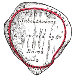
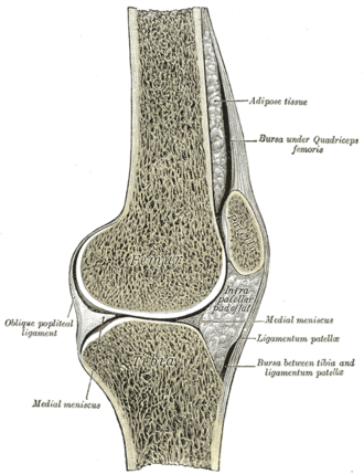
The prepatellar bursa lies between the skin and the patella; the infrapatellar fat pad lies behind the patellar ligament.
- Ossification
Several centers appear at about 3–6 years and fuse into one bone; failure of fusion leaves a bipartite patella (usually superolateral), which can mimic a fracture.
- Notes
The patella increases the moment arm of the quadriceps femoris. The Q angle (ASIS → center of patella → tibial tuberosity) is larger in women; a large Q angle and a shallow trochlear groove predispose to lateral patellar dislocation.
Muscle attachments
- Quadriceps femoris — insertion
Joints
Generated automatically from links in other entries.


Clinical correlations
Patellofemoral pain syndrome PANCE
- Mechanism / lesion
Overload of the patellofemoral joint, often with maltracking, quadriceps/hip weakness; the most common cause of anterior knee pain in young athletes (especially women).
- Classic presentation
Diffuse anterior knee pain worse with stairs, squatting, running and prolonged sitting ("theater sign"); treat with physical therapy (quadriceps and hip strengthening).
Lateral patellar dislocation PANCE
- Mechanism / lesion
Twisting on a planted foot with the knee slightly flexed; the patella dislocates laterally, often tearing the medial patellofemoral ligament.
- Classic presentation
Obvious lateral deformity (often reduced on extension); afterwards, effusion (hemarthrosis), medial tenderness and a positive apprehension test.
Patellar fracture
- Mechanism / lesion
A direct blow (fall onto the knee) or forceful quadriceps contraction.
- Classic presentation
Painful swelling and a palpable gap; inability to perform a straight-leg raise means the extensor mechanism is disrupted and usually needs surgery.
Prepatellar bursitis
- Mechanism / lesion
Repeated kneeling ("housemaid's knee") or infection (usually Staphylococcus aureus).
- Classic presentation
Swelling in front of the patella with preserved knee motion; aspirate if septic bursitis is suspected.
High-yield PANCE pearls
- Anterior knee pain with stairs and prolonged sitting → patellofemoral pain syndrome → PT.
- Patella dislocates laterally; positive apprehension test.
- Can't do a straight-leg raise after knee injury → extensor mechanism disruption (patellar fracture or tendon rupture).
Imaging anatomy
- X-ray
AP, lateral and sunrise (Merchant) view of the knee — fractures, patellar height (alta/baja) and tilt/subluxation. A bipartite patella has smooth, rounded margins.
- MRI
Shows MPFL tears, osteochondral fragments and the bone bruise pattern of a transient lateral dislocation (lateral femoral condyle and medial patella).
- Key views & landmarks
- Knee AP, lateral and sunrise (Merchant) views
Physical exam
- Surface landmarks
The patella, patellar ligament and tibial tuberosity are palpable in front of the knee.
- Palpation
Tenderness at the facets, poles (patellar or quadriceps tendinopathy) or medial retinaculum; check tracking during active flexion–extension.
- Strength / function testing
Straight-leg raise (integrity of the extensor mechanism).
Patellar apprehension test
- Technique
With the knee in about 20–30° of flexion, the examiner pushes the patella laterally.
- Positive test
The patient becomes apprehensive or contracts the quadriceps to stop the movement.
- Significance
Patellar instability or recent lateral dislocation.
Patellar grind (Clarke) test
- Technique
The examiner presses the patella down into the trochlea while the patient contracts the quadriceps.
- Positive test
Pain.
- Significance
Suggests patellofemoral pain, but many normal people are positive — low specificity.
Self-quiz
A 16-year-old girl has anterior knee pain that is worse going down stairs and after sitting through a movie. There is no effusion and ligament tests are normal. What is the most appropriate initial management?
Correct answer: C
This is patellofemoral pain syndrome. First-line treatment is physical therapy (quadriceps and hip abductor/external rotator strengthening) and activity modification; imaging is rarely needed.
Show answer
C
This is patellofemoral pain syndrome. First-line treatment is physical therapy (quadriceps and hip abductor/external rotator strengthening) and activity modification; imaging is rarely needed.
In which direction does the patella usually dislocate?
Correct answer: B
The patella almost always dislocates laterally, pulled by the valgus line of the quadriceps (Q angle) and limited by the medial patellofemoral ligament, which is usually torn.
Show answer
B
The patella almost always dislocates laterally, pulled by the valgus line of the quadriceps (Q angle) and limited by the medial patellofemoral ligament, which is usually torn.
After falling onto her knee, a patient has a palpable gap at the patella. Which finding shows the extensor mechanism is disrupted?
Correct answer: C
Inability to do a straight-leg raise means the extensor mechanism (quadriceps tendon, patella or patellar ligament) is disrupted, which usually needs surgery.
Show answer
C
Inability to do a straight-leg raise means the extensor mechanism (quadriceps tendon, patella or patellar ligament) is disrupted, which usually needs surgery.
A knee radiograph shows a smooth, well-corticated separate fragment at the superolateral corner of the patella in an athlete with no acute injury. What is the most likely explanation?
Correct answer: B
The patella ossifies from several centers; failure of fusion leaves a bipartite patella, usually superolateral, which can mimic a fracture. Smooth corticated edges favor a developmental variant.
Show answer
B
The patella ossifies from several centers; failure of fusion leaves a bipartite patella, usually superolateral, which can mimic a fracture. Smooth corticated edges favor a developmental variant.
A larger Q angle and a shallow femoral trochlear groove predispose to which injury?
Correct answer: D
The Q angle (ASIS → center of patella → tibial tuberosity) is larger in women. A large Q angle and a shallow trochlear groove predispose to lateral patellar dislocation, which often tears the medial patellofemoral ligament.
Show answer
D
The Q angle (ASIS → center of patella → tibial tuberosity) is larger in women. A large Q angle and a shallow trochlear groove predispose to lateral patellar dislocation, which often tears the medial patellofemoral ligament.