Spinal meninges and spaces
Three membranes (dura, arachnoid, pia) and the spaces between them surrounding the spinal cord. The epidural space is the target of epidural anesthesia, and the lumbar cistern is sampled by lumbar puncture.
Core anatomy
- Location
Within the vertebral canal, surrounding the spinal cord and cauda equina.
- Boundaries
- epidural space
- Between the dura and the walls of the canal (bone, ligamentum flavum). Contains fat and the internal vertebral venous plexus. Unlike the cranial epidural space, it is a real space
- dura mater
- Tough outer membrane; the dural sac ends at about S2
- subdural space
- Potential space between dura and arachnoid
- subarachnoid space
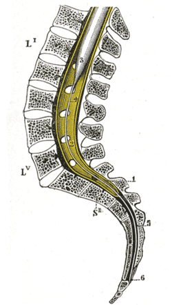
- Between arachnoid and pia; contains CSF. Below the conus it enlarges as the lumbar cistern (about L2–S2), holding the cauda equina
- pia mater
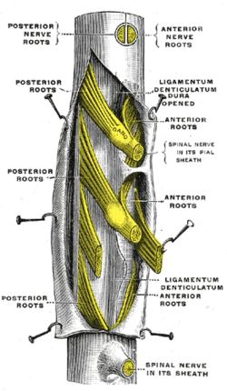
- Adheres to the cord; forms the denticulate ligaments (lateral anchors) and the filum terminale
- Contents
- CSF, cauda equina nerve roots, spinal vessels
- Communications
The subarachnoid space is continuous with the cranial subarachnoid space and ventricular system through the foramen magnum.
- Notes
Lumbar puncture is performed at L3–L4 or L4–L5 (identified by the intercristal line at L4), below the conus, where the needle pushes cauda equina roots aside rather than piercing the cord. Epidural anesthesia stops in the epidural space (loss of resistance after the ligamentum flavum); spinal anesthesia goes into the subarachnoid space.
Mentioned in
- Cauda equina
- Cranial meninges and intracranial hemorrhage
- Spinal cord
- Ventricular system and CSF
- Vertebral column
- Vertebral venous plexus
Generated automatically from links in other entries.
Swipe sideways to see the whole diagram →


Clinical correlations
Lumbar puncture indications, contraindications and CSF PANCE
- Mechanism / lesion
Sampling CSF from the lumbar cistern (meningitis, subarachnoid hemorrhage with a negative CT, demyelinating disease, pressure measurement).
- Classic presentation
Before LP, obtain head CT if there are signs of raised intracranial pressure or a mass (papilledema, focal deficits, altered consciousness, new seizure, immunocompromise). Other contraindications: coagulopathy or thrombocytopenia, skin infection at the site. In suspected bacterial meningitis, don't delay antibiotics for imaging or LP.
Post-dural puncture headache PANCE
- Mechanism / lesion
CSF leak through the dural hole lowers intracranial pressure.
- Classic presentation
Positional headache, worse sitting or standing and relieved lying flat, within days of LP or accidental dural puncture during epidural placement. Smaller, atraumatic (pencil-point) needles reduce the risk. Treatment: conservative care, caffeine, and an epidural blood patch if persistent.
Spinal epidural abscess PANCE
- Mechanism / lesion
Infection (usually Staphylococcus aureus) in the epidural space, via hematogenous spread (IV drug use, bacteremia) or procedures; risk factors include diabetes.
- Classic presentation
Classic triad of back pain, fever and neurologic deficit (often incomplete early). Emergent MRI with gadolinium, blood cultures, IV antibiotics and surgical decompression.
Spinal epidural hematoma
- Mechanism / lesion
Bleeding into the epidural space, especially with anticoagulation and neuraxial procedures (epidural catheter placement or removal).
- Classic presentation
Sudden back pain with rapidly progressive weakness or numbness and bladder dysfunction; emergency MRI and decompression.
High-yield PANCE pearls
- LP at L3–L4 or L4–L5 (intercristal line = L4).
- Get a head CT before LP if there are signs of raised ICP or focal deficits.
- Post-dural puncture headache: worse upright, better lying flat → epidural blood patch if persistent.
- Epidural abscess: fever + back pain + neurologic deficit, S. aureus, IV drug use → MRI with contrast.
- Epidural anesthesia stops in the epidural space; spinal anesthesia enters the subarachnoid space.
Imaging anatomy
- CT
Head CT before LP when indicated. CT myelography instills contrast into the subarachnoid space.
- MRI
MRI with gadolinium is the test of choice for epidural abscess, hematoma, and leptomeningeal disease; it also shows CSF leaks (with dedicated protocols).
- Ultrasound
Ultrasound-assisted LP identifies the midline and interspaces in obese patients or difficult anatomy.
- Key views & landmarks
- MRI spine with and without contrast
Physical exam
- Surface landmarks
Intercristal (Tuffier) line crossing L4 or the L4–L5 interspace; midline spinous processes.
- Palpation
For LP, position the patient in the lateral decubitus (fetal) position or sitting and flexed to open the interspaces; measure opening pressure only in the lateral position.
- Strength / function testing
Before LP, fundoscopy for papilledema and a focal neurologic exam. Afterward, monitor for headache and neurologic change.
Self-quiz
A 25-year-old woman develops a severe headache 2 days after a lumbar puncture. It is much worse when she sits up and improves when she lies down. What is the definitive treatment if conservative measures fail?
Correct answer: B
A positional post-dural puncture headache comes from a CSF leak. If conservative treatment fails, an epidural blood patch seals the dural hole.
Show answer
B
A positional post-dural puncture headache comes from a CSF leak. If conservative treatment fails, an epidural blood patch seals the dural hole.
A 40-year-old man who injects drugs has fever, severe thoracic back pain and new leg weakness. What is the most appropriate next step?
Correct answer: B
The triad of fever, back pain and neurologic deficit suggests a spinal epidural abscess. Obtain an emergent MRI with contrast, blood cultures, IV antibiotics, and surgical consultation.
Show answer
B
The triad of fever, back pain and neurologic deficit suggests a spinal epidural abscess. Obtain an emergent MRI with contrast, blood cultures, IV antibiotics, and surgical consultation.
Where does the needle tip lie in a correctly placed lumbar epidural (not spinal) anesthetic?
Correct answer: C
The epidural needle stops in the epidural space after the loss of resistance past the ligamentum flavum; puncturing the dura gives a "wet tap".
Show answer
C
The epidural needle stops in the epidural space after the loss of resistance past the ligamentum flavum; puncturing the dura gives a "wet tap".
The subarachnoid space (lumbar cistern) extends inferiorly to about which level?
Correct answer: D
The dural sac and subarachnoid space extend to about S2, well below the end of the cord (L1–L2).
Show answer
D
The dural sac and subarachnoid space extend to about S2, well below the end of the cord (L1–L2).
Before a lumbar puncture, which finding means a head CT should be done first?
Correct answer: B
Obtain a head CT before LP when there are signs of raised intracranial pressure or a mass: papilledema, focal deficits, altered consciousness, new seizure or immunocompromise. Don't delay antibiotics for imaging when bacterial meningitis is suspected.
Show answer
B
Obtain a head CT before LP when there are signs of raised intracranial pressure or a mass: papilledema, focal deficits, altered consciousness, new seizure or immunocompromise. Don't delay antibiotics for imaging when bacterial meningitis is suspected.