Internal jugular vein
Main venous drainage of the brain, face and neck. It runs in the carotid sheath and is the source of the jugular venous pressure (JVP), a common central line site, and the vein thrombosed in Lemierre syndrome.
Core anatomy
- Formation
Continuation of the sigmoid sinus at the jugular foramen (with CN IX, X and XI), dilated at its origin as the superior bulb.
- Course
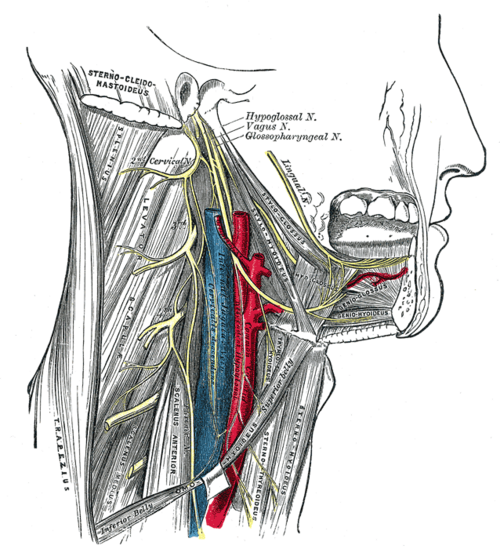
Descends in the carotid sheath, lateral to the internal and then common carotid (common and internal carotid arteries), with the vagus posterior between them, under cover of sternocleidomastoid. It lies deep to the gap between the sternal and clavicular heads of SCM low in the neck and has an inferior bulb with a valve just above its termination.
- Tributaries
- Inferior petrosal sinus
- Facial vein (via the common facial vein)
- Lingual and pharyngeal veins
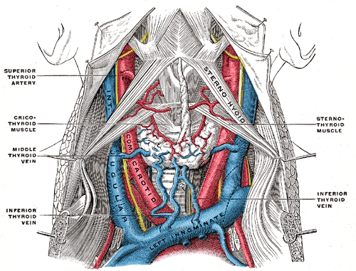
- Superior and middle thyroid veins (see thyroid gland)
- Drains into
Joins the subclavian vein behind the sternoclavicular joint to form the brachiocephalic vein → superior vena cava. The thoracic duct enters at the left venous angle (see thoracic duct).
- Relations
Deep cervical lymph nodes lie along the vein (see cervical lymph nodes); the right IJ runs in a nearly straight line to the right atrium.
- Notes
The right IJ is usually larger than the left and is preferred for central lines and for assessing the JVP.
Tributaries
Found in
Mentioned in
- Cervical lymph nodes
- Common and internal carotid arteries
- Sternocleidomastoid
- Thyroid gland
- Vagus nerve (CN X)
Generated automatically from links in other entries.


Clinical correlations
Jugular venous pressure (JVP) PANCE
- Mechanism / lesion
The right IJ runs in a straight line through the brachiocephalic vein and SVC to the right atrium, so its pulsation reflects right atrial pressure.
- Classic presentation
Measure the top of the IJ pulsation with the patient at 30–45°; normal is ≤ about 3–4 cm above the sternal angle (about 8 cm H₂O). Elevated in right heart failure, fluid overload, tricuspid regurgitation, cardiac tamponade, constrictive pericarditis and SVC obstruction (non-pulsatile). Kussmaul sign — JVP rises with inspiration (constrictive pericarditis). Hepatojugular reflux. verify
Central venous catheterization PANCE
- Mechanism / lesion
Ultrasound-guided cannulation, usually of the right IJ, at the apex of the triangle formed by the two heads of SCM and the clavicle, lateral to the carotid pulse.
- Classic presentation
Complications — carotid puncture, pneumothorax (less than the subclavian approach), air embolism (Trendelenburg position helps prevent it), infection, thrombosis. Confirm tip position with a chest X-ray (cavoatrial junction).
Lemierre syndrome PANCE
- Mechanism / lesion
Septic thrombophlebitis of the IJ after pharyngitis or peritonsillar abscess, classically Fusobacterium necrophorum; adolescents and young adults.
- Classic presentation
A recent sore throat, then high fever, lateral neck pain and swelling along SCM, and septic pulmonary emboli (cavitating nodules). Treat with IV antibiotics (e.g. ampicillin-sulbactam or a carbapenem).
High-yield PANCE pearls
- JVP is read on the right IJ at 30–45°; above ~3–4 cm over the sternal angle → elevated (right HF, tamponade, fluid overload).
- Kussmaul sign → constrictive pericarditis (also restrictive cardiomyopathy, right ventricular infarction; classically absent in tamponade).
- Pharyngitis → neck swelling → septic lung emboli → Lemierre syndrome (Fusobacterium).
- Central line → ultrasound guidance, then CXR to confirm tip and exclude pneumothorax.
Imaging anatomy
- CT
Contrast CT of the neck and chest shows IJ thrombosis (filling defect) and septic emboli in Lemierre syndrome.
- Ultrasound
Compressible, larger and lateral to the carotid; it distends with Valsalva. Non-compressible with echogenic contents → thrombosis.
- Key views & landmarks
- Point-of-care ultrasound for line placement
Physical exam
- Surface landmarks
Between the sternal and clavicular heads of SCM low in the neck; the sternal angle is the reference point for the JVP.
- Palpation
The IJ pulsation is not palpable (unlike the carotid) and is obliterated by light pressure above the clavicle.
- Strength / function testing
JVP: patient at 30–45°, head turned slightly left, tangential light; find the highest point of IJ pulsation and measure vertically from the sternal angle. Hepatojugular reflux: pressure over the liver for 10 seconds → a sustained rise of over 3 cm suggests right heart failure. The waveform is biphasic (a and v waves) and changes with position and respiration.
Self-quiz
Which feature best distinguishes the internal jugular venous pulsation from the carotid pulse on physical exam?
Correct answer: B
The jugular venous pulsation is not palpable, is obliterated by gentle pressure, has a biphasic waveform, falls with inspiration and changes with position. The carotid pulse is palpable and unaffected by these.
Show answer
B
The jugular venous pulsation is not palpable, is obliterated by gentle pressure, has a biphasic waveform, falls with inspiration and changes with position. The carotid pulse is palpable and unaffected by these.
A 19-year-old had a sore throat 1 week ago. He now has high fever, pain and swelling along the left sternocleidomastoid, and chest CT shows multiple cavitating nodules. What is the most likely diagnosis?
Correct answer: B
Lemierre syndrome — septic thrombophlebitis of the internal jugular vein after pharyngitis (classically Fusobacterium necrophorum) with septic pulmonary emboli.
Show answer
B
Lemierre syndrome — septic thrombophlebitis of the internal jugular vein after pharyngitis (classically Fusobacterium necrophorum) with septic pulmonary emboli.
A patient's jugular venous pressure rises during inspiration. Which condition does this Kussmaul sign most classically indicate?
Correct answer: A
Kussmaul sign (JVP rising with inspiration) classically indicates constrictive pericarditis (also restrictive cardiomyopathy and RV infarction). It is classically absent in tamponade.
Show answer
A
Kussmaul sign (JVP rising with inspiration) classically indicates constrictive pericarditis (also restrictive cardiomyopathy and RV infarction). It is classically absent in tamponade.
The internal jugular vein joins the subclavian vein to form the brachiocephalic vein behind which structure?
Correct answer: D
The IJ joins the subclavian vein behind the sternoclavicular joint to form the brachiocephalic vein. The thoracic duct enters at the left venous angle.
Show answer
D
The IJ joins the subclavian vein behind the sternoclavicular joint to form the brachiocephalic vein. The thoracic duct enters at the left venous angle.
During internal jugular central line placement, which patient position reduces the risk of air embolism?
Correct answer: C
The Trendelenburg position distends the neck veins and keeps their pressure above atmospheric, reducing the risk of air embolism. Other complications include carotid puncture, pneumothorax, infection and thrombosis.
Show answer
C
The Trendelenburg position distends the neck veins and keeps their pressure above atmospheric, reducing the risk of air embolism. Other complications include carotid puncture, pneumothorax, infection and thrombosis.