Pelvic diaphragm
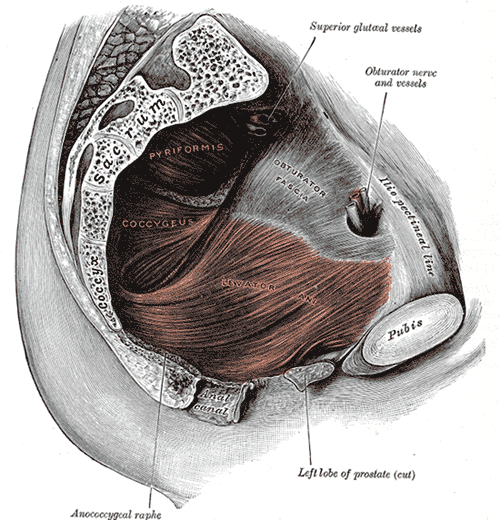
Funnel-shaped muscular floor of the pelvis formed by levator ani and coccygeus. It supports the pelvic organs and maintains urinary and fecal continence; its weakening underlies pelvic organ prolapse and stress incontinence.
Core anatomy
- Location
Spans the lesser pelvis between the pubis anteriorly, the lateral pelvic walls and the coccyx posteriorly, above the perineum and ischioanal fossae.
- Group
Muscles of the pelvic floor
- Origin
Levator ani: the body of the pubis, the tendinous arch of levator ani (a thickening of the obturator fascia), and the ischial spine. Coccygeus: the ischial spine and sacrospinous ligament.
- Insertion
Levator ani: the perineal body, the walls of the prostate or vagina, the rectum and anal canal, the anococcygeal body (ligament) and the coccyx. Coccygeus: the lateral margins of the lower sacrum and the coccyx.
- Innervation
Nerve to levator ani (direct branches of the ventral rami of S3–S4), with contributions from the inferior rectal and perineal branches of the pudendal nerve. Coccygeus: S3–S4 ventral rami. verify
- Blood supply
Branches of the inferior gluteal, inferior vesical and internal pudendal arteries (from the internal iliac artery).
- Action
- Supports the pelvic viscera and resists rises in intra-abdominal pressure (coughing, lifting, forced expiration)
- Puborectalis forms a U-shaped sling around the anorectal junction, maintaining the anorectal angle (fecal continence); it relaxes during defecation
- Supports the urethra and bladder neck (urinary continence), and contributes to vaginal closure
- Coccygeus supports the coccyx and pulls it forward after defecation or childbirth
- Relations
Parts of levator ani (medial to lateral): puborectalis, pubococcygeus (including puboprostaticus/ pubovaginalis and puboanalis fibers) and iliococcygeus. The urogenital hiatus between the medial borders transmits the urethra (and vagina); the rectum passes posteriorly. Above: the pelvic viscera and pelvic fascia. Below: the ischioanal fossae and the perineal membrane.
Mentioned in
Generated automatically from links in other entries.
Swipe sideways to see the whole diagram →

Clinical correlations
Pelvic organ prolapse PANCE
- Mechanism / lesion
Weakening or injury of levator ani and the connective-tissue supports (cardinal and uterosacral ligaments, pelvic fascia). Risk factors: vaginal delivery (especially operative or with a large baby), age and menopause, obesity, chronic cough or constipation, and heavy lifting.
- Classic presentation
Vaginal bulge or pressure, worse at the end of the day or with straining. Anterior wall (cystocele: incomplete emptying, UTIs), posterior wall (rectocele: need to splint the vagina to defecate), and apical (uterine or vault) prolapse.
- Notes
Graded with the POP-Q system. Management ranges from pelvic floor muscle training and a pessary to surgical repair.
Stress urinary incontinence PANCE
- Mechanism / lesion
Loss of urethral support (hypermobility) and/or weakness of the urethral sphincter (intrinsic sphincter deficiency), often after childbirth.
- Classic presentation
Leakage of small volumes with coughing, sneezing, laughing or lifting; no urgency. Positive cough stress test. First-line treatment is pelvic floor muscle (Kegel) training; midurethral sling surgery if that fails.
Levator ani avulsion
- Mechanism / lesion
Detachment of pubococcygeus/puborectalis from the pubis during vaginal delivery, especially forceps-assisted.
- Classic presentation
Widened urogenital hiatus on imaging; a strong risk factor for later prolapse.
High-yield PANCE pearls
- Kegel (pelvic floor) exercises are first-line for stress incontinence.
- Leak with cough/sneeze = stress incontinence; sudden urge then leak = urge incontinence (detrusor overactivity).
- Puborectalis sling maintains the anorectal angle for fecal continence.
- Prolapse risk factor most often tested: vaginal childbirth.
- Pelvic organ prolapse: pessary (non-surgical) vs surgical repair; grade with POP-Q.
Imaging anatomy
- MRI
Dynamic MRI (MR defecography) at rest and with straining shows descent of the bladder, cervix/vault and rectum relative to the pubococcygeal line, and levator defects.
- Ultrasound
Translabial (transperineal) 3-D/4-D ultrasound measures hiatal area and shows levator avulsion; used in urogynecology.
- Key views & landmarks
- Dynamic pelvic floor MRI at rest, squeeze and Valsalva
- Translabial ultrasound
Physical exam
- Surface landmarks
Levator ani is felt on digital vaginal exam as a muscular ridge along the lateral vaginal walls, and puborectalis as a sling posteriorly on digital rectal exam.
- Palpation
Ask the patient to squeeze around the examining finger: grade strength (e.g. modified Oxford scale 0–5), endurance and symmetry, and feel for a defect of the puborectalis near its pubic attachment.
- Strength / function testing
Inspect at rest and with Valsalva for prolapse (split speculum to see each vaginal wall separately); cough stress test with a comfortably full bladder.
Self-quiz
A 52-year-old woman who has had three vaginal deliveries reports leaking small amounts of urine when she coughs, laughs or lifts groceries. She has no urgency and no nocturia. What is the most appropriate first-line treatment?
Correct answer: C
This is stress urinary incontinence from weakened pelvic floor and urethral support. Pelvic floor muscle (Kegel) training is first-line; a midurethral sling is for failure of conservative therapy. Antimuscarinics and β3-agonists treat urge incontinence.
Show answer
C
This is stress urinary incontinence from weakened pelvic floor and urethral support. Pelvic floor muscle (Kegel) training is first-line; a midurethral sling is for failure of conservative therapy. Antimuscarinics and β3-agonists treat urge incontinence.
Which part of levator ani forms a sling around the anorectal junction and maintains the anorectal angle?
Correct answer: C
Puborectalis loops around the anorectal junction from the pubis, pulling it forward to create the anorectal angle. It relaxes during defecation to straighten the angle.
Show answer
C
Puborectalis loops around the anorectal junction from the pubis, pulling it forward to create the anorectal angle. It relaxes during defecation to straighten the angle.
Levator ani arises laterally from which structure?
Correct answer: B
Levator ani arises from the pubic body anteriorly, the tendinous arch of levator ani (a thickening of the obturator internus fascia) laterally, and the ischial spine posteriorly.
Show answer
B
Levator ani arises from the pubic body anteriorly, the tendinous arch of levator ani (a thickening of the obturator internus fascia) laterally, and the ischial spine posteriorly.
In a woman, which structures pass through the urogenital hiatus of levator ani?
Correct answer: B
The urogenital hiatus between the medial borders of levator ani transmits the urethra and vagina. The rectum passes through posteriorly, held by the puborectalis sling. A widened hiatus (e.g. after levator ani avulsion) is a risk factor for prolapse.
Show answer
B
The urogenital hiatus between the medial borders of levator ani transmits the urethra and vagina. The rectum passes through posteriorly, held by the puborectalis sling. A widened hiatus (e.g. after levator ani avulsion) is a risk factor for prolapse.
A 61-year-old woman with four vaginal deliveries has a vaginal bulge and must press on the posterior vaginal wall to complete a bowel movement. What is the most likely diagnosis?
Correct answer: E
A rectocele is a posterior vaginal wall prolapse; patients may need to splint the vagina to defecate. A cystocele (anterior wall) causes incomplete bladder emptying and UTIs. Vaginal delivery is the key risk factor; options include pelvic floor muscle training, a pessary or surgical repair.
Show answer
E
A rectocele is a posterior vaginal wall prolapse; patients may need to splint the vagina to defecate. A cystocele (anterior wall) causes incomplete bladder emptying and UTIs. Vaginal delivery is the key risk factor; options include pelvic floor muscle training, a pessary or surgical repair.