Triangle of auscultation and lumbar triangle
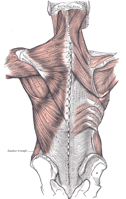
Two gaps in the superficial muscles of the back. The triangle of auscultation lets breath sounds be heard clearly; the lumbar (Petit) triangle is a rare site of lumbar hernia.
Core anatomy
- Location
Posterior thoracic wall (triangle of auscultation) and lower posterolateral abdominal wall just above the iliac crest (lumbar triangle).
- Boundaries
- auscultation — medial
- Lateral border of trapezius
- auscultation — inferior
- Superior border of latissimus dorsi
- auscultation — lateral
- Medial border of the scapula (with rhomboid major)
- auscultation — floor
- Thoracic wall (about the 6th–7th ribs and intercostal space), relatively free of overlying muscle
- lumbar (Petit) — inferior
- Iliac crest
- lumbar (Petit) — medial
- Lateral border of latissimus dorsi
- lumbar (Petit) — lateral
- Posterior border of the external oblique
- lumbar (Petit) — floor
- Internal oblique
- Contents
- No major neurovascular structures; each is simply a relatively thin area of the wall
- Notes
The triangle of auscultation enlarges when the scapula is drawn forward (arms folded across the chest and trunk flexed), improving access to breath sounds over the lower lobe posteriorly.

Clinical correlations
Auscultation of the lungs
- Mechanism / lesion
Thinner musculature over this area transmits breath sounds well.
- Classic presentation
Clear breath sounds from the lower lobe posteriorly. Historically also a surgical approach to the posterior thorax.
Lumbar (Petit) hernia
- Mechanism / lesion
Herniation through the inferior lumbar triangle (or the superior, Grynfeltt, triangle), congenital or acquired (trauma, surgery, iliac crest bone graft harvest).
- Classic presentation
A reducible bulge in the flank just above the iliac crest that enlarges with coughing; risk of incarceration.
High-yield PANCE pearls
- Triangle of auscultation: trapezius, latissimus dorsi, medial border of scapula; best heard with arms folded and trunk flexed.
- Lumbar (Petit) triangle: iliac crest, latissimus dorsi, external oblique → lumbar hernia.
Imaging anatomy
- CT
CT confirms lumbar hernia and its contents.
- Ultrasound
Dynamic ultrasound with Valsalva shows a lumbar hernia.
- Key views & landmarks
- CT abdomen for a flank bulge
Physical exam
- Surface landmarks
Triangle of auscultation near the inferior angle of the scapula, medial to it; lumbar triangle just above the iliac crest at the posterior axillary line region.
- Palpation
Palpate the flank for a bulge while the patient coughs (lumbar hernia).
- Strength / function testing
Auscultate posteriorly over the triangle with the patient's arms folded and trunk flexed forward.
Self-quiz
Which muscles bound the triangle of auscultation?
Correct answer: A
Trapezius (medial), latissimus dorsi (inferior) and the medial border of the scapula (lateral). Option B describes the lumbar triangle.
Show answer
A
Trapezius (medial), latissimus dorsi (inferior) and the medial border of the scapula (lateral). Option B describes the lumbar triangle.
How can the triangle of auscultation be enlarged during the examination?
Correct answer: B
Folding the arms and flexing the trunk protracts the scapula, widening the triangle.
Show answer
B
Folding the arms and flexing the trunk protracts the scapula, widening the triangle.
A flank bulge just above the iliac crest between latissimus dorsi and external oblique is most likely a hernia through which area?
Correct answer: B
The inferior lumbar (Petit) triangle is bounded by the iliac crest, latissimus dorsi and external oblique.
Show answer
B
The inferior lumbar (Petit) triangle is bounded by the iliac crest, latissimus dorsi and external oblique.