Parathyroid glands
Usually four small endocrine glands on the back of the thyroid that secrete parathyroid hormone. They are important in hypercalcemia (primary hyperparathyroidism) and in hypocalcemia after thyroid surgery.
Core anatomy
- Location
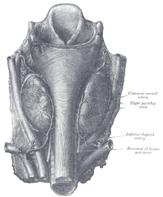
On the posterior surface of the thyroid lobes, usually two on each side (superior and inferior); each is about 5 mm, yellow-brown.
- Parts
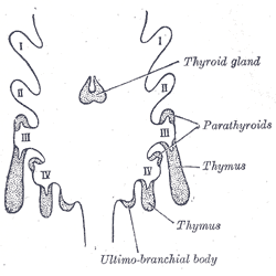
- Superior parathyroids: from the 4th pharyngeal pouch; fairly constant position near the cricothyroid junction
- Inferior parathyroids: from the 3rd pharyngeal pouch (with the thymus); descend further and are more variable — may be in the thymus or anterior mediastinum (see thymus)
- Chief cells secrete parathyroid hormone (PTH) — raises serum calcium (bone resorption, renal calcium reabsorption and phosphate excretion, activation of vitamin D)
- Number varies — supernumerary or fewer than four glands in a minority of people
- Relations
Close to the recurrent laryngeal nerve and the inferior thyroid artery behind the thyroid gland.
- Arterial supply
Mainly the inferior thyroid artery (both superior and inferior glands in most people).
- Venous drainage
Thyroid venous plexus.
- Lymphatic drainage
With the thyroid (see cervical lymph nodes).
- Innervation
Sympathetic vasomotor fibers; secretion is controlled by serum ionized calcium via the calcium-sensing receptor.
Found in
Mentioned in
Generated automatically from links in other entries.


Clinical correlations
Primary hyperparathyroidism PANCE
- Mechanism / lesion
Single parathyroid adenoma (about 80–85%), four-gland hyperplasia (MEN 1, MEN 2A), rarely carcinoma.
- Classic presentation
Often asymptomatic hypercalcemia on routine labs; otherwise "stones, bones, groans, thrones and psychiatric overtones" — kidney stones, osteoporosis, abdominal pain/peptic ulcer/pancreatitis, polyuria, depression. Labs: high calcium with high or inappropriately normal PTH, low phosphate. Treatment: parathyroidectomy when indicated.
Secondary and tertiary hyperparathyroidism PANCE
- Mechanism / lesion
Secondary — a physiological response to low calcium, most often chronic kidney disease or vitamin D deficiency. Tertiary — autonomous PTH secretion after long-standing secondary disease.
- Classic presentation
Secondary: high PTH, low or normal calcium, high phosphate in CKD. Tertiary: high PTH with high calcium.
Hypoparathyroidism PANCE
- Mechanism / lesion
Most often after thyroid or parathyroid surgery (gland removal or devascularization); also autoimmune, DiGeorge syndrome (absent 3rd/4th pouches), hypomagnesemia.
- Classic presentation
Hypocalcemia: perioral and fingertip tingling, cramps, Chvostek sign (facial twitch on tapping the facial nerve), Trousseau sign (carpal spasm with a BP cuff), prolonged QT, seizures, laryngospasm. Treat symptomatic cases with IV calcium gluconate.
High-yield PANCE pearls
- High calcium + high/normal PTH → primary hyperparathyroidism (usually a single adenoma).
- "Stones, bones, groans, thrones, psychiatric overtones."
- Tingling and carpal spasm after thyroidectomy → hypocalcemia → Chvostek/Trousseau → IV calcium gluconate.
- CKD → secondary hyperparathyroidism (high PTH, high phosphate).
- Inferior parathyroids = 3rd pouch (with thymus); superior = 4th pouch.
Imaging anatomy
- CT
4D-CT of the neck for localization before minimally invasive parathyroidectomy.
- Ultrasound
Neck ultrasound to localize an adenoma (after the biochemical diagnosis).
- Key views & landmarks
- Sestamibi (Tc-99m) scan — adenoma retains tracer
- Imaging localizes; it does not diagnose hyperparathyroidism
Physical exam
- Surface landmarks
Not palpable normally — a palpable neck mass with hypercalcemia raises concern for parathyroid carcinoma.
- Strength / function testing
Chvostek sign: tap the facial nerve in front of the ear → twitch of the upper lip. Trousseau sign: inflate a BP cuff above systolic for 3 minutes → carpal spasm (more specific).
Self-quiz
A 58-year-old woman has a calcium of 11.2 mg/dL on routine labs, a low phosphate and an elevated PTH. She has had a kidney stone. What is the most likely cause?
Correct answer: B
High calcium with elevated PTH means PTH-dependent hypercalcemia; most cases are primary hyperparathyroidism from a single adenoma. Malignancy (PTHrP) suppresses PTH. Check urine calcium to exclude familial hypocalciuric hypercalcemia.
Show answer
B
High calcium with elevated PTH means PTH-dependent hypercalcemia; most cases are primary hyperparathyroidism from a single adenoma. Malignancy (PTHrP) suppresses PTH. Check urine calcium to exclude familial hypocalciuric hypercalcemia.
Two days after total thyroidectomy, a patient reports tingling around the mouth. Tapping in front of the ear causes the upper lip to twitch. What is the best treatment?
Correct answer: B
Perioral tingling and a positive Chvostek sign indicate hypocalcemia from parathyroid injury or removal. Symptomatic hypocalcemia is treated with IV calcium gluconate, then oral calcium and calcitriol.
Show answer
B
Perioral tingling and a positive Chvostek sign indicate hypocalcemia from parathyroid injury or removal. Symptomatic hypocalcemia is treated with IV calcium gluconate, then oral calcium and calcitriol.
The inferior parathyroid glands may be found in the anterior mediastinum. Why?
Correct answer: C
The inferior parathyroids arise from the 3rd pouch with the thymus and descend further, so they are more variable and may lie in the thymus or anterior mediastinum. The superior glands arise from the 4th pouch and are more constant.
Show answer
C
The inferior parathyroids arise from the 3rd pouch with the thymus and descend further, so they are more variable and may lie in the thymus or anterior mediastinum. The superior glands arise from the 4th pouch and are more constant.
Which artery is the main blood supply of both the superior and inferior parathyroid glands in most people?
Correct answer: D
The inferior thyroid artery usually supplies both parathyroids on each side, so preserving it during thyroidectomy reduces postoperative hypoparathyroidism.
Show answer
D
The inferior thyroid artery usually supplies both parathyroids on each side, so preserving it during thyroidectomy reduces postoperative hypoparathyroidism.
A patient on dialysis for CKD has which pattern of labs in secondary hyperparathyroidism?
Correct answer: B
In secondary hyperparathyroidism of CKD, PTH rises in response to low calcium and phosphate retention: high PTH, low/normal calcium, high phosphate. Primary disease gives high calcium with low phosphate; tertiary gives high PTH with high calcium.
Show answer
B
In secondary hyperparathyroidism of CKD, PTH rises in response to low calcium and phosphate retention: high PTH, low/normal calcium, high phosphate. Primary disease gives high calcium with low phosphate; tertiary gives high PTH with high calcium.