Thoracic duct
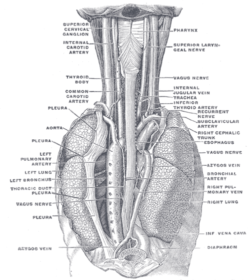
Largest lymphatic vessel. It drains lymph from most of the body, ascends through the posterior mediastinum, and empties into the left venous angle. Injury causes chylothorax.
Core anatomy
- Location
Posterior mediastinum, between the thoracic aorta and the azygos vein, behind the esophagus.
- Drains
Lymph (and absorbed dietary fat as chyle) from the entire body except the right upper quadrant: the right side of the head and neck, the right upper limb, and the right side of the thorax drain to the right lymphatic duct.
- Drains to
The left venous angle, the junction of the left internal jugular and left subclavian veins.
- Notes
Course: begins at the cisterna chyli (at about L1–L2) in the abdomen. It passes through the aortic hiatus (T12) of the diaphragm, ascends in the posterior mediastinum between the thoracic aorta (left) and the azygos vein (right), posterior to the esophagus. At about T4–T6 it crosses to the left and ascends along the left side of the esophagus into the neck. There it arches laterally (at about C7) to open into the left venous angle. verify
Found in
Mentioned in
- Azygos vein
- Cervical lymph nodes
- Esophagus
- Internal jugular vein
- Pelvic and inguinal lymph drainage
- Pleural cavity
- Thoracic aorta
Generated automatically from links in other entries.

Clinical correlations
Chylothorax PANCE
- Mechanism / lesion
Leakage of chyle into the pleural cavity from injury to the duct (thoracic or esophageal surgery, trauma, central line placement in the left neck) or obstruction by malignancy (lymphoma is a classic cause). Injury below the crossing point usually causes a right chylothorax; injury above it, a left chylothorax.
- Classic presentation
Pleural effusion with milky white fluid. Triglycerides above 110 mg/dL (and chylomicrons) confirm it. Prolonged leaks cause malnutrition, lymphopenia and immunosuppression.
Virchow node PANCE
- Mechanism / lesion
Metastatic spread (classically gastric and other abdominal cancers) along the thoracic duct to the left supraclavicular node near its termination.
- Classic presentation
A hard, enlarged left supraclavicular lymph node (Troisier sign).
High-yield PANCE pearls
- Thoracic duct: cisterna chyli (L1–L2) → aortic hiatus (T12) → crosses to the left (~T5) → left venous angle.
- Chylothorax: milky pleural fluid, triglycerides > 110 mg/dL; after thoracic surgery, trauma, or lymphoma.
- Virchow node (left supraclavicular) → think gastric/abdominal malignancy.
- Drains everything except the right head/neck, right arm, and right thorax (right lymphatic duct).
Imaging anatomy
- X-ray
Pleural effusion (not specific for chyle).
- CT
Effusion and possible causes (mediastinal lymphoma, post-surgical changes). Fat attenuation of the fluid is not reliable.
- MRI
MR lymphangiography can map the duct and localize leaks.
- Key views & landmarks
- Thoracentesis with triglyceride and chylomicron measurement (the diagnostic test)
- Lymphangiography (conventional or MR) to localize a leak and guide embolization
Physical exam
- Surface landmarks
Terminates near the junction of the left internal jugular and subclavian veins, behind the left sternoclavicular joint area.
- Palpation
Palpate the left supraclavicular fossa for a Virchow node.
- Strength / function testing
Effusion signs on the chest exam (dullness, decreased fremitus and breath sounds).
Self-quiz
Five days after an esophagectomy, a patient has an enlarging right pleural effusion. Thoracentesis yields milky white fluid with a triglyceride level of 350 mg/dL. Which structure was injured?
Correct answer: B
Chylothorax after esophageal surgery comes from thoracic duct injury. Below its crossing point the duct lies on the right, so lower injuries produce a right-sided chylothorax.
Show answer
B
Chylothorax after esophageal surgery comes from thoracic duct injury. Below its crossing point the duct lies on the right, so lower injuries produce a right-sided chylothorax.
Where does the thoracic duct usually empty?
Correct answer: B
The thoracic duct ends at the left venous angle. The right lymphatic duct drains into the right venous angle.
Show answer
B
The thoracic duct ends at the left venous angle. The right lymphatic duct drains into the right venous angle.
A 70-year-old man with weight loss and early satiety has a hard, enlarged left supraclavicular lymph node. Which malignancy should be suspected?
Correct answer: B
A Virchow node (left supraclavicular) reflects spread of abdominal malignancy, classically gastric cancer, via the thoracic duct.
Show answer
B
A Virchow node (left supraclavicular) reflects spread of abdominal malignancy, classically gastric cancer, via the thoracic duct.
Where does the thoracic duct begin, and through which diaphragmatic opening does it enter the thorax?
Correct answer: C
The thoracic duct begins at the cisterna chyli (about L1–L2), passes through the aortic hiatus (T12), ascends between the aorta and azygos vein, crosses to the left and empties into the left venous angle.
Show answer
C
The thoracic duct begins at the cisterna chyli (about L1–L2), passes through the aortic hiatus (T12), ascends between the aorta and azygos vein, crosses to the left and empties into the left venous angle.
Which region drains to the right lymphatic duct rather than the thoracic duct?
Correct answer: A
The right lymphatic duct drains the right side of the head and neck, the right upper limb and the right side of the thorax. The thoracic duct drains everything else, including the whole body below the diaphragm.
Show answer
A
The right lymphatic duct drains the right side of the head and neck, the right upper limb and the right side of the thorax. The thoracic duct drains everything else, including the whole body below the diaphragm.