Oculomotor nerve (CN III)
Supplies four extraocular muscles, levator palpebrae and the pupillary constrictor. A palsy gives a "down and out" eye with ptosis; a blown pupil signals compression (aneurysm or herniation) rather than diabetic ischemia.
Core anatomy
- Origin
Oculomotor nucleus (somatic motor) and Edinger–Westphal nucleus (parasympathetic) in the midbrain at the level of the superior colliculus. The nerve emerges in the interpeduncular fossa, medial to the cerebral peduncle.
- Course
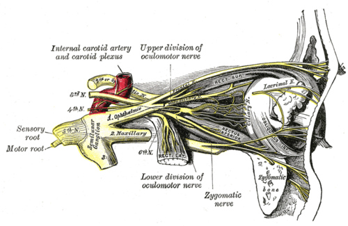
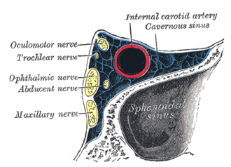
Passes between the posterior cerebral and superior cerebellar arteries, runs forward beside the posterior communicating artery, crosses the edge of the tentorium near the uncus, then runs in the lateral wall of the cavernous sinus (see dural venous sinuses) and enters the orbit through the superior orbital fissure within the common tendinous ring, dividing into superior and inferior divisions.
- Branches
- Superior division: superior rectus, levator palpebrae superioris
- Inferior division: medial rectus, inferior rectus, inferior oblique, and the parasympathetic root to the ciliary ganglion
- Motor
Extraocular muscles — superior, inferior and medial recti and inferior oblique; levator palpebrae superioris. Parasympathetic (via the ciliary ganglion and short ciliary nerves): sphincter pupillae (miosis) and ciliary muscle (accommodation).
- Relations
The parasympathetic fibers lie superficially (dorsomedial) on the nerve, so compression affects the pupil early, while ischemia (vasa nervorum, core of the nerve) tends to spare it.
Other structures innervated
Found in
Mentioned in
Generated automatically from links in other entries.
Swipe sideways to see the whole diagram →


Clinical correlations
Third nerve palsy PANCE
- Mechanism / lesion
Compressive: posterior communicating artery aneurysm, uncal (transtentorial) herniation, tumor. Ischemic (microvascular): diabetes, hypertension. Also midbrain stroke, cavernous sinus lesions, trauma.
- Classic presentation
Eye "down and out" (unopposed lateral rectus and superior oblique), ptosis, and — if the parasympathetic fibers are involved — a fixed dilated pupil and loss of accommodation; diplopia. Pupil-involving palsy (especially painful, sudden) = aneurysm until proven otherwise → emergency CTA/MRA. Pupil-sparing palsy in an older diabetic = usually microvascular ischemia, recovers over about 3 months.
Uncal herniation PANCE
- Mechanism / lesion
A supratentorial mass (e.g. epidural or subdural hematoma) pushes the medial temporal lobe (uncus) over the tentorium, compressing CN III.
- Classic presentation
Ipsilateral blown pupil followed by third nerve palsy, decreasing consciousness, and contralateral hemiparesis (or ipsilateral from Kernohan notch compression of the opposite peduncle). Neurosurgical emergency.
Weber syndrome
- Mechanism / lesion
Paramedian midbrain infarct (branches of the PCA) involving the CN III fascicles and the cerebral peduncle.
- Classic presentation
Ipsilateral CN III palsy with contralateral hemiparesis.
High-yield PANCE pearls
- "Down and out" eye + ptosis → CN III palsy.
- CN III palsy with a dilated pupil → PComm aneurysm (or herniation) → emergency CTA.
- Pupil-sparing CN III palsy in a diabetic → microvascular ischemia.
- Unilateral blown pupil in a head-injured patient → uncal herniation.
- Ipsilateral CN III palsy + contralateral hemiparesis → Weber syndrome (midbrain).
Imaging anatomy
- CT
Noncontrast CT head for trauma/herniation; CT angiography for suspected PComm aneurysm.
- MRI
MRI with MRA to evaluate the midbrain, cavernous sinus and aneurysm; catheter angiography if noninvasive imaging is negative but suspicion is high.
Physical exam
- Strength / function testing
Inspect for ptosis and eye position at rest; H-pattern eye movements (adduction, elevation, depression); pupil size, direct and consensual light reflex and accommodation (near response). See the H-test diagram on extraocular muscles.
Self-quiz
A 52-year-old woman has a sudden severe headache and drooping of the left eyelid. The left eye is deviated down and out, and the left pupil is 7 mm and unreactive. What is the most likely cause?
Correct answer: B
A pupil-involving third nerve palsy with sudden headache indicates compression of the superficial parasympathetic fibers, classically by a posterior communicating artery aneurysm — obtain emergency CTA.
Show answer
B
A pupil-involving third nerve palsy with sudden headache indicates compression of the superficial parasympathetic fibers, classically by a posterior communicating artery aneurysm — obtain emergency CTA.
A 68-year-old man with poorly controlled diabetes has double vision and right ptosis. The right eye is down and out; the pupils are equal and reactive. What is the most likely mechanism?
Correct answer: B
A pupil-sparing CN III palsy in a patient with vascular risk factors is usually microvascular ischemia, which affects the core of the nerve and spares the superficial pupillary fibers. It typically recovers within about 3 months.
Show answer
B
A pupil-sparing CN III palsy in a patient with vascular risk factors is usually microvascular ischemia, which affects the core of the nerve and spares the superficial pupillary fibers. It typically recovers within about 3 months.
Why does a compressive lesion of CN III affect the pupil early, while ischemic lesions often spare it?
Correct answer: C
The parasympathetic fibers lie on the surface of the nerve, so external compression (aneurysm, herniation) affects the pupil early, while microvascular ischemia of the nerve's core tends to spare it.
Show answer
C
The parasympathetic fibers lie on the surface of the nerve, so external compression (aneurysm, herniation) affects the pupil early, while microvascular ischemia of the nerve's core tends to spare it.
Between which two arteries does the oculomotor nerve emerge?
Correct answer: E
CN III emerges from the interpeduncular fossa and passes between the posterior cerebral and superior cerebellar arteries, then runs alongside the posterior communicating artery.
Show answer
E
CN III emerges from the interpeduncular fossa and passes between the posterior cerebral and superior cerebellar arteries, then runs alongside the posterior communicating artery.
A patient with a large left subdural hematoma has a blown left pupil but weakness on the LEFT side. What explains the ipsilateral weakness?
Correct answer: B
In uncal herniation, the midbrain can be pushed so that the opposite cerebral peduncle is compressed against the tentorial edge (Kernohan notch), causing weakness on the same side as the lesion, a false localizing sign.
Show answer
B
In uncal herniation, the midbrain can be pushed so that the opposite cerebral peduncle is compressed against the tentorial edge (Kernohan notch), causing weakness on the same side as the lesion, a false localizing sign.
Flashcards
See also
- Extraocular muscles
- Midbrain
- Circle of Willis
- Dural venous sinuses
- Orbit
- Cranial nerves at a glance (comparison table)
- A lucid interval after a blow to the head (clinical case)