Supraspinatus
Rotator cuff muscle in the supraspinous fossa that initiates abduction and is the most commonly torn cuff tendon.
Core anatomy
- Location
Supraspinous fossa of the scapula, passing under the acromion to the top of the humeral head.
- Group
Rotator cuff (SITS = Supraspinatus, Infraspinatus, Teres minor, Subscapularis); scapulohumeral muscles.
- Origin
Supraspinous fossa of the scapula and the deep fascia covering it.
- Insertion
Superior facet of the greater tubercle of the humerus.
- Innervation
Suprascapular nerve (C5, C6) verify
- Blood supply
Suprascapular artery verify
- Action
- Initiates abduction of the arm (roughly the first 15°); assists deltoid through the rest of abduction
- With the other rotator cuff muscles, holds the humeral head in the glenoid cavity
- Relations
The tendon passes beneath the coracoacromial arch (acromion, coracoacromial ligament, coracoid process). The subacromial (subdeltoid) bursa separates it from the arch and deltoid. It blends with the superior capsule of the glenohumeral joint. The suprascapular nerve reaches the muscle by passing through the suprascapular notch beneath the superior transverse scapular ligament.
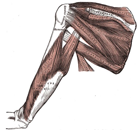
Swipe sideways to see the whole diagram →

Clinical correlations
Rotator cuff tear PANCE
- Mechanism / lesion
Supraspinatus is the most commonly torn cuff tendon. Tears are usually degenerative in patients over ~40 (attrition under the arch, reduced vascularity near the insertion) and are sometimes acute (fall, lifting, or dislocation in older patients).
- Classic presentation
Lateral shoulder pain, often at night and when lying on the affected side. Weak abduction and external rotation. Full-thickness tears may not allow the patient to lower the arm smoothly from abduction (drop-arm sign). Passive ROM is typically preserved, unlike adhesive capsulitis.
Subacromial impingement / rotator cuff tendinopathy PANCE
- Mechanism / lesion
Irritation of the supraspinatus tendon and subacromial bursa beneath the coracoacromial arch with repetitive overhead activity (painters, swimmers, throwers). Contributors include acromial shape, AC osteophytes and poor scapular mechanics.
- Classic presentation
A painful arc between about 60° and 120° of abduction. Pain with overhead activity. Positive Neer and Hawkins–Kennedy signs. Pain with the empty-can test, with preserved strength if there's no tear.
Calcific tendinitis
- Mechanism / lesion
Deposition of calcium hydroxyapatite within the tendon, most often supraspinatus.
- Classic presentation
Acute, severe shoulder pain with markedly restricted motion during the resorptive phase; calcification visible on X-ray.
High-yield PANCE pearls
- Most commonly torn rotator cuff tendon: supraspinatus.
- Empty-can (Jobe) test isolates supraspinatus; drop-arm test suggests a full-thickness cuff tear.
- Painful arc (60–120°) and Neer/Hawkins signs suggest subacromial impingement.
- Cuff pathology: passive ROM preserved, active ROM limited. Adhesive capsulitis: both active and passive ROM limited.
- MRI (or ultrasound) is used to confirm and grade rotator cuff tears.
Imaging anatomy
- X-ray
Often normal. Look for calcific tendinitis (calcification above the greater tuberosity), a hooked acromion or AC osteophytes (outlet view), and changes in the greater tuberosity. In chronic massive tears the humeral head rides up and the acromiohumeral interval narrows. verify
- MRI
Oblique coronal T2 fat-saturated images show the tendon from the muscle belly to the footprint. A full-thickness tear appears as a fluid-bright gap through the full tendon thickness, with or without retraction. Oblique sagittal images assess fatty infiltration and atrophy of the muscle belly, which predicts outcome after repair. MR arthrography: contrast leaking into the subacromial–subdeltoid bursa indicates a full-thickness tear.
- Ultrasound
Normal tendon: a hyperechoic, fibrillar "beak" inserting on the greater tuberosity beneath the deltoid and bursa, best seen with the arm extended and internally rotated (hand in back pocket). Full-thickness tear: a hypo- or anechoic defect, non-visualization of the tendon, or loss of the normal convex bursal contour. Dynamic scanning can show impingement.
- Key views & landmarks
- Shoulder AP (internal and external rotation), scapular Y/outlet, axillary
- MRI oblique coronal and oblique sagittal T2 fat-sat
- Ultrasound long and short axis with the arm in the modified Crass (hand-in-back-pocket) position
Physical exam
- Surface landmarks
The muscle belly lies in the supraspinous fossa above the spine of the scapula (atrophy appears as hollowing). The tendon insertion can be palpated just anterior/inferior to the anterolateral acromion when the shoulder is extended.
- Palpation
Extend and internally rotate the arm (hand behind the back) to bring the insertion out from under the acromion, then palpate for tenderness below the anterolateral acromion.
- Strength / function testing
Resisted abduction in the scapular plane. Inspect for supraspinous fossa atrophy.
Empty-can (Jobe) test
- Technique
Arms abducted to 90° in the scapular plane (about 30° anterior to the coronal plane) and fully internally rotated (thumbs pointing down). The patient resists downward pressure.
- Positive test
Pain and/or weakness compared with the other side.
- Significance
Supraspinatus tendinopathy (pain) or tear (weakness).
Drop-arm test
- Technique
The examiner passively abducts the arm to about 90°, and the patient slowly lowers it to the side.
- Positive test
The arm drops suddenly or can't be lowered smoothly.
- Significance
Suggests a full-thickness rotator cuff tear.
Painful arc
- Technique
The patient actively abducts the arm through its full range.
- Positive test
Pain between about 60° and 120°, easing above and below that range.
- Significance
Subacromial impingement / cuff tendinopathy.
Neer and Hawkins–Kennedy impingement signs
- Technique
Neer: stabilize the scapula and passively forward-flex the internally rotated arm fully. Hawkins–Kennedy: forward-flex the shoulder and elbow to 90°, then passively internally rotate the shoulder.
- Positive test
Pain in the anterolateral shoulder.
- Significance
Subacromial impingement.
Self-quiz
A 58-year-old house painter has 4 months of right lateral shoulder pain that wakes him at night. Pain occurs between 70° and 110° of active abduction. Abduction against resistance with the arm internally rotated and the thumb down is painful and weak. Passive range of motion is full. Which muscle is most likely involved?
Correct answer: B
A painful arc and a painful, weak empty-can test indicate supraspinatus tendinopathy or tear. Full passive ROM argues against adhesive capsulitis. Infraspinatus and teres minor are tested by resisted external rotation, subscapularis by lift-off/belly-press.
Show answer
B
A painful arc and a painful, weak empty-can test indicate supraspinatus tendinopathy or tear. Full passive ROM argues against adhesive capsulitis. Infraspinatus and teres minor are tested by resisted external rotation, subscapularis by lift-off/belly-press.
Supraspinatus is innervated by a nerve arising from which part of the brachial plexus?
Correct answer: B
The suprascapular nerve arises from the superior trunk (C5–C6) and supplies supraspinatus and infraspinatus. It passes through the suprascapular notch below the superior transverse scapular ligament.
Show answer
B
The suprascapular nerve arises from the superior trunk (C5–C6) and supplies supraspinatus and infraspinatus. It passes through the suprascapular notch below the superior transverse scapular ligament.
An examiner passively abducts a patient's arm to 90° and asks her to lower it slowly. The arm falls abruptly to her side. What is the most likely diagnosis?
Correct answer: B
A positive drop-arm test suggests a full-thickness rotator cuff tear, usually involving supraspinatus. Adhesive capsulitis limits passive motion rather than causing the arm to drop.
Show answer
B
A positive drop-arm test suggests a full-thickness rotator cuff tear, usually involving supraspinatus. Adhesive capsulitis limits passive motion rather than causing the arm to drop.
A patient with a complete supraspinatus tear would have the most difficulty with which movement?
Correct answer: A
Supraspinatus initiates abduction (about the first 15°) and keeps the humeral head seated so deltoid can continue abduction. Patients often shrug or lean to start the movement.
Show answer
A
Supraspinatus initiates abduction (about the first 15°) and keeps the humeral head seated so deltoid can continue abduction. Patients often shrug or lean to start the movement.
Which finding best distinguishes a rotator cuff tear from adhesive capsulitis?
Correct answer: E
In rotator cuff pathology passive range of motion is preserved while active motion is limited. In adhesive capsulitis both active and passive motion are lost, most markedly external rotation. Night pain and pain with overhead activity occur in both.
Show answer
E
In rotator cuff pathology passive range of motion is preserved while active motion is limited. In adhesive capsulitis both active and passive motion are lost, most markedly external rotation. Night pain and pain with overhead activity occur in both.