Gluteus medius and minimus
Fan-shaped hip abductors that keep the pelvis level when standing on one leg. Weakness or superior gluteal nerve injury produces the Trendelenburg sign; their tendons cause lateral hip pain.
Core anatomy
- Location
Deep to gluteus maximus (medius) and deep to medius (minimus), on the outer surface of the ilium.
- Group
Gluteal region, intermediate and deep layers (abductors and medial rotators)
- Origin
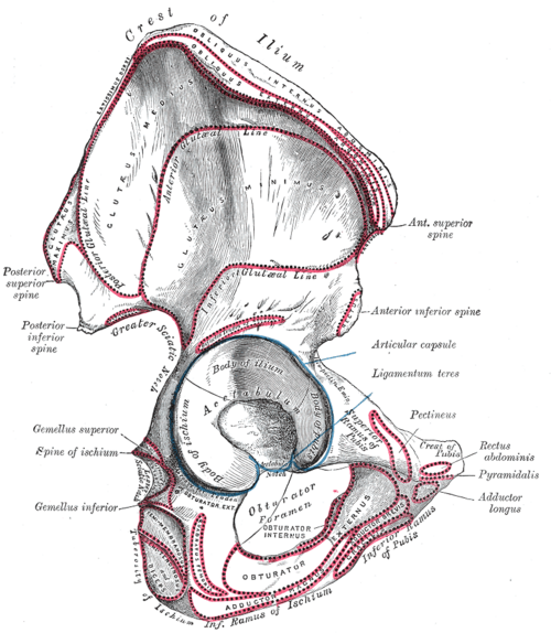
External surface of the ilium of the hip bone — medius between the anterior and posterior gluteal lines; minimus between the anterior and inferior gluteal lines.
- Insertion
Greater trochanter of the femur — medius on its lateral surface; minimus on its anterior surface.
- Innervation
Superior gluteal nerve (L4–S1), from the sacral plexus
- Blood supply
Superior gluteal artery (from the internal iliac artery).
- Action
- Abduction of the hip
- Stabilize the pelvis on the stance limb during walking, keeping the opposite side from dropping
- Medial rotation of the hip (anterior fibers)
- Relations
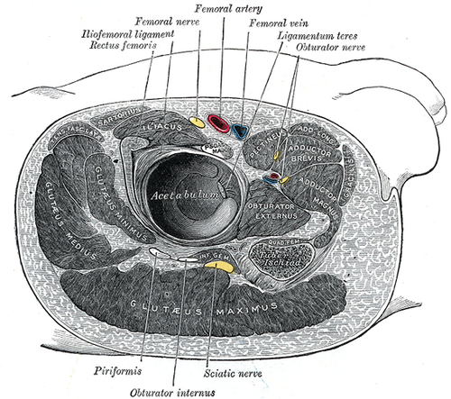
The superior gluteal nerve and vessels run between the two muscles; tensor fasciae latae (same nerve) lies anteriorly.


Clinical correlations
Trendelenburg sign and gait PANCE
- Mechanism / lesion
Weakness of gluteus medius and minimus — superior gluteal nerve injury (injection, hip surgery), L5 radiculopathy, hip OA, DDH or a fractured greater trochanter.
- Classic presentation
When standing on the affected leg, the opposite side of the pelvis drops (positive Trendelenburg sign). Walking, the patient lurches the trunk toward the affected side (compensated Trendelenburg/"waddling" gait if bilateral).
Gluteal tendinopathy (greater trochanteric pain syndrome) PANCE
- Mechanism / lesion
Degeneration or tears of the medius/minimus tendons at the greater trochanter, often with bursitis; common in middle-aged women.
- Classic presentation
Lateral hip pain, trochanteric tenderness, pain lying on the affected side or standing on one leg; treat with load management, physical therapy (abductor strengthening) and sometimes injection.
High-yield PANCE pearls
- Positive Trendelenburg = weak gluteus medius/minimus (superior gluteal nerve, L5) on the stance side; the opposite hip drops.
- Lateral hip pain with trochanteric tenderness → gluteal tendinopathy/bursitis.
Imaging anatomy
- MRI
Gluteal tendon tears and tendinopathy; denervation edema after nerve injury.
- Ultrasound
Tendinopathy, tears, bursitis; guides injection.
Physical exam
- Surface landmarks
The muscles lie between the iliac crest and the greater trochanter on the lateral hip.
- Palpation
Tenderness at the greater trochanter.
- Strength / function testing
Resisted hip abduction in side-lying (hip slightly extended); observe gait.
Trendelenburg test
- Technique
The patient stands on the tested leg and lifts the other foot off the ground for about 30 seconds.
- Positive test
The pelvis drops on the side of the lifted leg (or the trunk leans toward the stance side).
- Significance
Weak hip abductors on the stance side (superior gluteal nerve, L5 root, hip joint disease).
Self-quiz
After an intramuscular injection into the right buttock, a patient's left hip drops whenever she stands on her right leg. Which muscle is weak?
Correct answer: B
When standing on the right leg, the right gluteus medius and minimus hold the pelvis level. If they are weak (right superior gluteal nerve injury), the unsupported left side drops.
Show answer
B
When standing on the right leg, the right gluteus medius and minimus hold the pelvis level. If they are weak (right superior gluteal nerve injury), the unsupported left side drops.
Which nerve root lesion most commonly weakens the hip abductors?
Correct answer: D
Hip abduction is mainly L5 (superior gluteal nerve L4–S1). An L5 radiculopathy can cause a Trendelenburg sign along with weak great toe extension and ankle dorsiflexion.
Show answer
D
Hip abduction is mainly L5 (superior gluteal nerve L4–S1). An L5 radiculopathy can cause a Trendelenburg sign along with weak great toe extension and ankle dorsiflexion.
Which nerve supplies gluteus medius and gluteus minimus?
Correct answer: C
The superior gluteal nerve (L4–S1) runs between gluteus medius and minimus and also supplies tensor fasciae latae. The inferior gluteal nerve supplies gluteus maximus.
Show answer
C
The superior gluteal nerve (L4–S1) runs between gluteus medius and minimus and also supplies tensor fasciae latae. The inferior gluteal nerve supplies gluteus maximus.
A patient with a weak left gluteus medius walks by lurching the trunk sideways each time the left foot is on the ground. Toward which side does the trunk lurch?
Correct answer: A
Leaning the trunk toward the weak stance side moves the center of gravity over the hip, reducing the abductor force needed. This is the compensated Trendelenburg gait. The uncompensated sign is the opposite side of the pelvis dropping.
Show answer
A
Leaning the trunk toward the weak stance side moves the center of gravity over the hip, reducing the abductor force needed. This is the compensated Trendelenburg gait. The uncompensated sign is the opposite side of the pelvis dropping.
A 52-year-old woman has lateral hip pain that is worse when she lies on that side or stands on one leg. There is tenderness over the greater trochanter, and groin pain and hip rotation are normal. What is the most likely diagnosis?
Correct answer: D
Gluteal tendinopathy (greater trochanteric pain syndrome) affects the medius and minimus tendons at the greater trochanter, often with bursitis, and is common in middle-aged women. Hip OA typically causes groin pain with loss of internal rotation.
Show answer
D
Gluteal tendinopathy (greater trochanteric pain syndrome) affects the medius and minimus tendons at the greater trochanter, often with bursitis, and is common in middle-aged women. Hip OA typically causes groin pain with loss of internal rotation.
Flashcards
See also
- Gluteus maximus
- Hip joint
- Sacral plexus
- Lower limb nerve injuries (comparison table)