Knee joint
The largest synovial joint — a modified hinge between the femur, tibia and patella, stabilized mainly by ligaments and the menisci. Ligament and meniscal injuries, effusions and osteoarthritis make it one of the most examined joints.
Core anatomy
- Location
Between the distal femur, the proximal tibia and the patella; the popliteal fossa lies behind it.
- Classification
Synovial modified hinge (bicondylar) joint, with a patellofemoral (plane/saddle) component
- Articulating surfaces
Femur (condyles), tibia (plateau) and patella; the fibula is not part of the joint
- Capsule
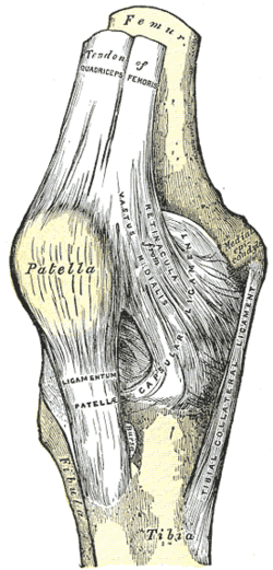
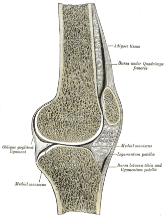
Thin and incomplete anteriorly, where the quadriceps tendon, patella and patellar ligament take its place. The synovial membrane forms the suprapatellar bursa, which communicates with the joint (why effusions fill above the patella).
- Ligaments
- Cruciate ligaments (ACL and PCL): intra-articular, extrasynovial; anteroposterior stability
- Collateral ligaments of the knee (MCL and LCL): varus/valgus stability
- Patellar ligament: continuation of the quadriceps tendon to the tibial tuberosity
- Oblique and arcuate popliteal ligaments (posterior capsule)
- Menisci of the knee: medial (C-shaped, attached to the MCL) and lateral (O-shaped, more mobile)
- Movements
- Flexion (hamstrings, gastrocnemius, popliteus)
- Extension (quadriceps femoris)
- Slight medial/lateral rotation of the flexed knee
- Screw-home mechanism: the femur rotates medially on the tibia in the last degrees of extension, locking the knee; popliteus unlocks it by rotating the femur laterally
- Stability
Mostly ligamentous and muscular (quadriceps), since the bony surfaces are poorly congruent.
- Innervation
Branches of the femoral nerve, obturator nerve, tibial nerve and common fibular nerve (genicular nerves).
- Blood supply
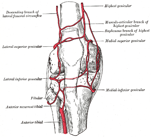
Genicular anastomosis — genicular branches of the popliteal artery, the descending genicular branch of the femoral artery, the descending branch of the lateral circumflex femoral, and the anterior tibial recurrent artery.
- Relations
Bursae around the knee include prepatellar, infrapatellar, pes anserine (medial tibia) and semimembranosus bursae; a popliteal (Baker) cyst is a distended gastrocnemius–semimembranosus bursa.
Mentioned in
Generated automatically from links in other entries.
Swipe sideways to see the whole diagram →



Clinical correlations
Knee effusion and hemarthrosis PANCE
- Mechanism / lesion
Acute hemarthrosis within hours of injury suggests ACL tear (most common), patellar dislocation or intra-articular fracture. Non-bloody effusions: OA, inflammatory or crystal arthritis, infection.
- Classic presentation
Swelling filling the suprapatellar pouch and obscuring the "dimples" beside the patella. Aspirate an unexplained effusion (cell count, Gram stain, culture, crystals).
Knee osteoarthritis PANCE
- Mechanism / lesion
Degenerative cartilage loss, most often in the medial compartment (varus deformity).
- Classic presentation
Activity-related pain, short morning stiffness, crepitus, bony enlargement; X-ray with joint space narrowing and osteophytes. Treat with exercise, weight loss, topical then oral NSAIDs, intra-articular steroids, then total knee arthroplasty.
Septic arthritis and crystal arthritis PANCE
- Mechanism / lesion
The knee is the most commonly affected joint in both septic arthritis (S. aureus; gonococcus in young sexually active adults) and pseudogout (CPPD).
- Classic presentation
Acute hot, swollen knee — aspirate. Synovial WBC over about 50,000/µL suggests infection; rhomboid, positively birefringent crystals = CPPD; needle-shaped negatively birefringent = gout.
Popliteal (Baker) cyst
- Mechanism / lesion
Fluid from the joint (OA, meniscal tears, RA) distends the gastrocnemius–semimembranosus bursa.
- Classic presentation
Posterior knee swelling; rupture causes calf pain and swelling that mimics DVT (crescent sign) — Doppler ultrasound distinguishes them.
High-yield PANCE pearls
- Rapid hemarthrosis after a pivoting injury → ACL tear.
- Hot swollen knee → aspirate (septic vs crystals).
- Rhomboid, positively birefringent crystals = pseudogout; knee is the most common site.
- Ruptured Baker cyst mimics DVT → ultrasound.
- Popliteus unlocks the knee; the femur rotates medially to lock it (screw-home).
Imaging anatomy
- X-ray
AP (weight-bearing for OA), lateral, and sunrise views. Ottawa knee rules decide who needs films after trauma. Chondrocalcinosis in CPPD; fat–fluid level in intra-articular fractures; Segond fracture (lateral tibial avulsion) implies an ACL tear.
- MRI
The test of choice for ligament, meniscal and cartilage injuries and bone bruises.
- Ultrasound
Effusions, Baker cysts, tendon injuries; guides aspiration.
- Key views & landmarks
- Weight-bearing AP (standing) for joint-space narrowing
- MRI knee without contrast
Physical exam
- Surface landmarks
The joint line is palpable on each side of the patellar ligament with the knee flexed; the tibial tuberosity, fibular head, femoral epicondyles and pes anserinus below the medial joint line.
- Palpation
Joint-line tenderness (meniscus), MCL/LCL tenderness, effusion: bulge (wipe) test for small effusions and the patellar tap (ballottement) for larger ones.
- Strength / function testing
Inspect alignment (varus/valgus), swelling and quadriceps bulk; range of motion (full extension 0°, flexion about 135°); gait. Ligament and meniscal tests — see cruciate ligaments, collateral ligaments of the knee and menisci of the knee. Patellar reflex (L4).
Bulge (wipe) test
- Technique
Stroke fluid up out of the medial recess into the suprapatellar pouch, then stroke down the lateral side.
- Positive test
A bulge of fluid reappears on the medial side.
- Significance
Detects small effusions.
Self-quiz
A 72-year-old woman has a hot, swollen right knee. Aspiration shows 25,000 WBC/µL and rhomboid-shaped, weakly positively birefringent crystals. X-ray shows linear calcification of the menisci. What is the diagnosis?
Correct answer: B
Rhomboid, positively birefringent crystals with chondrocalcinosis indicate calcium pyrophosphate deposition (pseudogout), which most often affects the knee. Gout crystals are needle-shaped and negatively birefringent.
Show answer
B
Rhomboid, positively birefringent crystals with chondrocalcinosis indicate calcium pyrophosphate deposition (pseudogout), which most often affects the knee. Gout crystals are needle-shaped and negatively birefringent.
Which muscle "unlocks" the fully extended knee at the start of flexion?
Correct answer: B
Popliteus rotates the femur laterally on the fixed tibia (or the tibia medially when the foot is free), reversing the screw-home locking so flexion can begin.
Show answer
B
Popliteus rotates the femur laterally on the fixed tibia (or the tibia medially when the foot is free), reversing the screw-home locking so flexion can begin.
A 55-year-old man with knee OA has sudden calf pain and swelling after gardening. Homans sign is equivocal. What is the best initial test?
Correct answer: B
A ruptured Baker cyst and DVT look alike. Compression (duplex) ultrasound excludes DVT and can show the cyst. Treat for DVT only if a thrombus is seen.
Show answer
B
A ruptured Baker cyst and DVT look alike. Compression (duplex) ultrasound excludes DVT and can show the cyst. Treat for DVT only if a thrombus is seen.
Knee osteoarthritis most often affects which compartment, producing which deformity?
Correct answer: D
Degenerative cartilage loss in the knee is most common in the medial compartment, producing a varus (bow-legged) deformity.
Show answer
D
Degenerative cartilage loss in the knee is most common in the medial compartment, producing a varus (bow-legged) deformity.
Why does a knee effusion cause swelling above the patella?
Correct answer: E
The synovial membrane forms the suprapatellar bursa, which communicates with the joint, so effusions fill the space above the patella and obscure the dimples beside it. The prepatellar bursa lies in front of the patella and does not communicate with the joint.
Show answer
E
The synovial membrane forms the suprapatellar bursa, which communicates with the joint, so effusions fill the space above the patella and obscure the dimples beside it. The prepatellar bursa lies in front of the patella and does not communicate with the joint.