Anterior interventricular artery
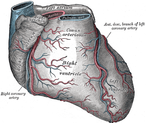
Branch of the left coronary artery in the anterior interventricular groove. It supplies the anterior LV wall, the anterior two-thirds of the septum and the apex, and is the most commonly occluded coronary artery.
Core anatomy
- Location
Anterior interventricular groove, from the left main bifurcation around the apex.
- Origin
- Course
Descends in the anterior interventricular groove toward the apex, often curving around it onto the inferior surface.
- Branches
- Diagonal branches: to the anterolateral LV wall
- Septal perforating branches: to the anterior two-thirds of the interventricular septum (including the bundle branches)
- Branches to the anterolateral papillary muscle (shared with the circumflex)
- Supplies
Anterior wall of the left ventricle, the anterior two-thirds of the interventricular septum, the apex, part of the anterior right ventricle, and (with the circumflex) the anterolateral papillary muscle.
- Anastomoses
At the apex with the posterior interventricular artery.
Structures supplied
Mentioned in
Generated automatically from links in other entries.

Clinical correlations
Anterior (anteroseptal) myocardial infarction PANCE
- Mechanism / lesion
LAD occlusion. Proximal occlusion (the "widowmaker") endangers a large area of myocardium.
- Classic presentation
ST elevation in V1–V4 (septal V1–V2, anterior V3–V4), with extension to V5–V6, I and aVL for anterolateral involvement. Complications: heart failure/cardiogenic shock, new bundle branch block, LV apical aneurysm and thrombus, and ventricular septal rupture.
Wellens syndrome PANCE
- Mechanism / lesion
Critical proximal LAD stenosis in a patient whose pain has resolved.
- Classic presentation
Deeply inverted or biphasic T waves in V2–V3 in a pain-free patient. High risk of an extensive anterior MI; needs urgent angiography, and stress testing is contraindicated.
CABG target PANCE
- Mechanism / lesion
Surgical bypass of LAD stenosis.
- Classic presentation
The LIMA → LAD graft (internal thoracic artery) has the best long-term patency.
High-yield PANCE pearls
- LAD = most commonly occluded coronary artery; anterior MI V1–V4.
- Proximal LAD = "widowmaker" (large territory at risk).
- Wellens: biphasic/deep T inversions V2–V3 → critical LAD stenosis; no stress test.
- LIMA–LAD is the preferred bypass graft.
Imaging anatomy
- CT
Coronary CTA shows stenosis and plaque. LV apical aneurysm or thrombus may be visible.
- MRI
Late gadolinium enhancement in the anterior wall, septum and apex.
- Ultrasound
Echo shows anterior, septal and apical hypokinesis/akinesis, reduced EF, apical aneurysm or LV thrombus.
- Key views & landmarks
- Invasive angiography (RAO cranial and LAO cranial views show the LAD and diagonals)
- 12-lead ECG: V1–V4
Physical exam
- Strength / function testing
Assess for heart failure (S3, crackles, JVD) and a new holosystolic murmur (ventricular septal rupture) after anterior MI.
Self-quiz
A 58-year-old woman has chest pain with ST elevation in V1–V4. Which artery is most likely occluded?
Correct answer: C
V1–V4 = anteroseptal wall = LAD territory. Inferior (II, III, aVF) = RCA; lateral (I, aVL, V5–V6) = circumflex.
Show answer
C
V1–V4 = anteroseptal wall = LAD territory. Inferior (II, III, aVF) = RCA; lateral (I, aVL, V5–V6) = circumflex.
Which region of the heart receives its blood supply mainly from the LAD?
Correct answer: B
The septal perforators of the LAD supply the anterior two-thirds of the septum. The posterior third comes from the posterior interventricular artery.
Show answer
B
The septal perforators of the LAD supply the anterior two-thirds of the septum. The posterior third comes from the posterior interventricular artery.
A pain-free 55-year-old man admitted after an episode of chest pain has deeply inverted T waves in V2 and V3 with normal troponin. What is the most appropriate next step?
Correct answer: C
This is Wellens syndrome, a marker of critical proximal LAD stenosis. Stress testing may precipitate an MI; the patient needs coronary angiography.
Show answer
C
This is Wellens syndrome, a marker of critical proximal LAD stenosis. Stress testing may precipitate an MI; the patient needs coronary angiography.
Several weeks after a large anterior MI, echocardiography shows a thin, dyskinetic apex containing a mural thrombus. What complication has developed?
Correct answer: E
The LAD supplies the apex, so a large anterior MI can leave a left ventricular apical aneurysm, a thin dyskinetic segment in which thrombus forms (embolic risk). Papillary muscle rupture typically follows an inferior MI.
Show answer
E
The LAD supplies the apex, so a large anterior MI can leave a left ventricular apical aneurysm, a thin dyskinetic segment in which thrombus forms (embolic risk). Papillary muscle rupture typically follows an inferior MI.
Which bypass conduit to the LAD has the best long-term patency?
Correct answer: A
The LIMA → LAD graft has the best long-term patency of any bypass conduit and is the standard graft to the LAD in CABG.
Show answer
A
The LIMA → LAD graft has the best long-term patency of any bypass conduit and is the standard graft to the LAD in CABG.