Clavicle
S-shaped strut connecting the upper limb to the axial skeleton; one of the most commonly fractured bones, usually in its middle third.
Core anatomy
- Location
Subcutaneous along its whole length at the root of the neck, from the manubrium to the acromion.
- Classification
Long bone that lies horizontally. It is described as lacking a true medullary cavity (spongy bone within a compact shell). verify
- Key features & landmarks
- Sternal (medial) end: enlarged, articulates with the manubrium at the sternoclavicular joint
- Acromial (lateral) end: flat, articulates with the acromion at the acromioclavicular joint
- Medial two-thirds convex anteriorly; lateral third flattened and concave anteriorly
- Conoid tubercle and trapezoid line (inferior surface, lateral third): attachments of the coracoclavicular ligament
- Impression for the costoclavicular ligament (inferior surface, medial end)
- Subclavian groove (inferior surface, middle third): attachment of subclavius
- Relations
The subclavian vessels and the trunks and divisions of the brachial plexus pass posterior and inferior to the middle third, protected by subclavius. It acts as a strut holding the limb away from the trunk and transmits force from the upper limb to the axial skeleton.
- Ossification
The first bone to begin ossifying (around weeks 5–6 of gestation), largely by intramembranous ossification. The medial (sternal) epiphysis is among the last to fuse, in the early-to-mid 20s. verify
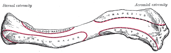
Muscle attachments
- Deltoid — origin
- Pectoralis major — origin
- Sternocleidomastoid — origin
- Trapezius — insertion
Forms a boundary of
Mentioned in
Generated automatically from links in other entries.

Clinical correlations
Clavicle fracture PANCE
- Mechanism / lesion
Fall onto the shoulder or on an outstretched hand. The middle third is the most common site (roughly 80%). Also occurs in newborns during difficult deliveries (shoulder dystocia).
- Classic presentation
Pain, swelling and a visible or palpable deformity. The patient supports the arm. The medial fragment is pulled superiorly by sternocleidomastoid, and the lateral fragment is pulled down by the weight of the limb and drawn medially by pectoralis major. Check the skin (tenting) and the neurovascular status of the limb.
Neonatal clavicle fracture
- Mechanism / lesion
Birth trauma, particularly with shoulder dystocia or large birth weight.
- Classic presentation
Crepitus or a palpable lump, decreased movement of the arm, and an asymmetric Moro reflex. Must be distinguished from Erb palsy, which can coexist.
Cleidocranial dysplasia
- Mechanism / lesion
Autosomal dominant disorder (RUNX2) affecting intramembranous ossification.
- Classic presentation
Hypoplastic or absent clavicles (the shoulders can be brought together in front of the chest), delayed closure of the fontanelles, and dental anomalies.
High-yield PANCE pearls
- Most common location of clavicle fracture: middle third.
- Medial fragment displaced up (SCM); lateral fragment displaced down (weight of the arm).
- Most midshaft clavicle fractures are managed non-operatively with a sling; open fractures, skin tenting, neurovascular injury or marked displacement/shortening prompt orthopedic referral.
- Clavicle fracture in a newborn → asymmetric Moro reflex; check for coexisting brachial plexus injury.
Imaging anatomy
- X-ray
AP clavicle plus an AP view with 15–30° cephalic tilt, which projects the clavicle clear of the ribs and scapula to show displacement. Assess shortening and displacement. Include the AC and SC joints.
- CT
CT is preferred for medial clavicle and sternoclavicular joint injuries (especially posterior SC dislocation, which can compress the mediastinal great vessels or trachea) and for surgical planning.
- Ultrasound
Can diagnose neonatal clavicle fractures without radiation.
- Key views & landmarks
- AP clavicle
- AP with 15–30° cephalic tilt
- CT for sternoclavicular joint
Physical exam
- Surface landmarks
Palpable along its whole length from the jugular notch to the acromion. Its lateral end forms a small step above the acromion at the AC joint.
- Palpation
Palpate from the SC joint to the AC joint for step-off, crepitus and point tenderness. Check the skin over any deformity.
- Strength / function testing
Assess distal pulses and brachial plexus function (motor and sensory) in any clavicle injury.
Self-quiz
A 19-year-old cyclist falls onto his left shoulder. There is a deformity in the middle of the clavicle. Which displacement is expected?
Correct answer: B
Sternocleidomastoid pulls the medial fragment up. The weight of the limb (and pectoralis major) pulls the lateral fragment down and medially.
Show answer
B
Sternocleidomastoid pulls the medial fragment up. The weight of the limb (and pectoralis major) pulls the lateral fragment down and medially.
Which structures lie immediately posterior/inferior to the middle third of the clavicle and are at risk in a fracture?
Correct answer: B
The subclavian artery and vein and the trunks and divisions of the brachial plexus pass behind the middle third of the clavicle, separated from it by subclavius.
Show answer
B
The subclavian artery and vein and the trunks and divisions of the brachial plexus pass behind the middle third of the clavicle, separated from it by subclavius.
Which radiographic view best shows displacement of a midshaft clavicle fracture in addition to the standard AP?
Correct answer: B
The cephalic-tilt AP view projects the clavicle above the ribs and scapula, showing superior displacement and shortening. The scapular Y and axillary views are for the glenohumeral joint.
Show answer
B
The cephalic-tilt AP view projects the clavicle above the ribs and scapula, showing superior displacement and shortening. The scapular Y and axillary views are for the glenohumeral joint.
A child can bring both shoulders together in front of the chest. He also has delayed closure of the fontanelles and dental anomalies. Which process is primarily affected?
Correct answer: B
Cleidocranial dysplasia is an autosomal dominant RUNX2 disorder of intramembranous ossification, giving hypoplastic or absent clavicles, delayed fontanelle closure and dental anomalies. FGFR3 mutations cause achondroplasia; type I collagen defects cause osteogenesis imperfecta.
Show answer
B
Cleidocranial dysplasia is an autosomal dominant RUNX2 disorder of intramembranous ossification, giving hypoplastic or absent clavicles, delayed fontanelle closure and dental anomalies. FGFR3 mutations cause achondroplasia; type I collagen defects cause osteogenesis imperfecta.
A 24-year-old falls onto his shoulder and has a closed, minimally displaced midshaft clavicle fracture. The skin is intact and the neurovascular exam is normal. What is the most appropriate management?
Correct answer: C
Most midshaft clavicle fractures are managed non-operatively with a sling. Open fractures, skin tenting, neurovascular injury or marked displacement/shortening prompt orthopedic referral.
Show answer
C
Most midshaft clavicle fractures are managed non-operatively with a sling. Open fractures, skin tenting, neurovascular injury or marked displacement/shortening prompt orthopedic referral.