Intervertebral disc
Fibrocartilaginous joint between vertebral bodies, made of a tough anulus fibrosus around a gel-like nucleus pulposus. Lumbar disc herniation at L4–L5 or L5–S1 causes sciatica.
Core anatomy
- Location
Between adjacent vertebral bodies from C2–C3 to L5–S1 (none between C1 and C2).
- Classification
Secondary cartilaginous joint (symphysis).
- Articulating surfaces
Vertebral bodies of adjacent vertebrae, covered by cartilaginous end plates.
- Capsule
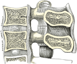
None (not synovial). The anulus fibrosus (concentric lamellae of fibrocartilage) encloses the nucleus pulposus (gelatinous, a remnant of the notochord), which acts as a shock absorber.
- Ligaments
- Anterior longitudinal ligament (strong, anterior)
- Posterior longitudinal ligament (weaker and narrower, especially in the lumbar region, so herniations go posterolateral)
- Movements
- Small movements at each level that add up to the spine's overall flexion, extension, lateral flexion and rotation
- Stability
Anulus fibrosus and longitudinal ligaments.
- Innervation
The outer anulus is innervated by the sinuvertebral (recurrent meningeal) nerves; the inner disc is aneural and largely avascular (nourished by diffusion), so it heals poorly.
- Notes
Which root is compressed? In the lumbar spine a typical posterolateral (paracentral) herniation compresses the traversing root, the one numbered for the lower vertebra (L4–L5 → L5, L5–S1 → S1). A far-lateral (foraminal) herniation compresses the exiting root, numbered for the upper vertebra (L4–L5 → L4). In the cervical spine a C5–C6 herniation compresses C6. A large central herniation can compress the cauda equina.
Swipe sideways to see the whole diagram →

Clinical correlations
Lumbar disc herniation (sciatica) PANCE
- Mechanism / lesion
Degeneration and tears of the anulus allow the nucleus to herniate, usually posterolaterally at L4–L5 or L5–S1, often after lifting or flexion–rotation. Peak age 30–50.
- Classic presentation
Low back pain with radicular leg pain below the knee, worse with sitting, coughing or sneezing. Dermatomal numbness, myotomal weakness and reflex changes. Positive straight leg raise (and crossed SLR, which is more specific). L4: knee extension, patellar reflex, medial leg. L5: great-toe extension and ankle dorsiflexion, hip abduction, dorsum of the foot, no reliable reflex. S1: plantarflexion and eversion, Achilles reflex, lateral foot and sole.
Red flags in low back pain PANCE
- Mechanism / lesion
Features that suggest fracture, cancer, infection or cauda equina syndrome rather than a mechanical cause.
- Classic presentation
Age over 50 with new pain, cancer history, unexplained weight loss, fever or IV drug use, immunosuppression, significant trauma or osteoporosis, night pain or pain at rest, progressive neurologic deficit, saddle anesthesia and urinary retention or incontinence. Without red flags, acute low back pain is managed without imaging (stay active, NSAIDs, heat), since most improve within about 6 weeks.
High-yield PANCE pearls
- L4–L5 posterolateral herniation → L5 root; L5–S1 → S1 root; far-lateral L4–L5 → L4.
- L4: patellar reflex, knee extension. L5: great-toe extension/dorsiflexion, no reflex. S1: Achilles reflex, plantarflexion.
- Straight leg raise (30–70°) is sensitive; crossed SLR is specific for herniation.
- No red flags → no imaging for acute low back pain; MRI if red flags or persistent deficits after about 6 weeks of conservative care.
Imaging anatomy
- X-ray
Not useful for disc herniation (discs are radiolucent). Disc space narrowing and osteophytes indicate degeneration.
- CT
CT myelography if MRI is contraindicated.
- MRI
MRI is the test of choice: herniation size and location, root compression, and red-flag pathology. Many asymptomatic people have disc bulges, so correlate with the exam.
- Key views & landmarks
- MRI lumbar spine (sagittal and axial T2)
Physical exam
- Surface landmarks
Iliac crests at L4 help identify the L4–L5 interspace.
- Strength / function testing
Heel walking (L5, dorsiflexion) and toe walking (S1, plantarflexion). Great-toe extension (L5), knee extension (L4). Patellar (L4) and Achilles (S1) reflexes. Dermatomal sensation. Check perianal sensation and ask about bladder function when cauda equina is a concern.
Straight leg raise (Lasègue)
- Technique
With the patient supine and the knee straight, passively raise the leg.
- Positive test
Radicular pain radiating below the knee between about 30° and 70° of elevation (back or hamstring pain alone doesn't count). Dorsiflexing the ankle may increase it.
- Significance
L5 or S1 nerve root tension (disc herniation). Sensitive but not specific.
Crossed straight leg raise
- Technique
Raise the asymptomatic leg.
- Positive test
Pain in the symptomatic leg.
- Significance
Highly specific for lumbar disc herniation.
Self-quiz
A 38-year-old man has back pain radiating down the lateral leg to the dorsum of the foot after lifting a heavy box. He has weak great-toe extension and normal patellar and Achilles reflexes. A posterolateral herniation at which level is most likely?
Correct answer: C
Weak great-toe extension and dorsum-of-foot numbness with normal reflexes indicate an L5 radiculopathy. A posterolateral herniation at L4–L5 compresses the traversing L5 root.
Show answer
C
Weak great-toe extension and dorsum-of-foot numbness with normal reflexes indicate an L5 radiculopathy. A posterolateral herniation at L4–L5 compresses the traversing L5 root.
A patient has sciatica with weak plantarflexion, numbness of the lateral foot, and an absent ankle reflex. Which root is affected?
Correct answer: D
S1: plantarflexion (gastrocnemius–soleus), lateral foot and sole sensation, and the Achilles reflex.
Show answer
D
S1: plantarflexion (gastrocnemius–soleus), lateral foot and sole sensation, and the Achilles reflex.
A 34-year-old woman has 1 week of mechanical low back pain without radiation, trauma or systemic symptoms. Neurologic exam is normal. What is the most appropriate management?
Correct answer: C
Acute low back pain without red flags is managed conservatively without imaging. Staying active is better than bed rest; most patients improve within weeks.
Show answer
C
Acute low back pain without red flags is managed conservatively without imaging. Staying active is better than bed rest; most patients improve within weeks.
Why do lumbar disc herniations usually occur posterolaterally?
Correct answer: B
The posterior longitudinal ligament is narrow in the lumbar region, leaving the posterolateral anulus relatively unsupported.
Show answer
B
The posterior longitudinal ligament is narrow in the lumbar region, leaving the posterolateral anulus relatively unsupported.
A far-lateral (foraminal) disc herniation at L4–L5 compresses which nerve root?
Correct answer: E
A typical posterolateral L4–L5 herniation compresses the traversing L5 root, but a far-lateral (foraminal) herniation compresses the exiting root, numbered for the upper vertebra: L4.
Show answer
E
A typical posterolateral L4–L5 herniation compresses the traversing L5 root, but a far-lateral (foraminal) herniation compresses the exiting root, numbered for the upper vertebra: L4.