Vertebrobasilar system
The posterior circulation — the vertebral arteries join to form the basilar artery, which supplies the brainstem, cerebellum and (through the PCAs) the occipital lobes. Its strokes cause vertigo, crossed signs and, with basilar occlusion, coma or locked-in syndrome.
Core anatomy
- Origin
Vertebral arteries (vertebral artery), first branches of the subclavian arteries, ascending through the transverse foramina of C6–C1 and entering the skull through the foramen magnum.
- Course
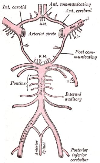
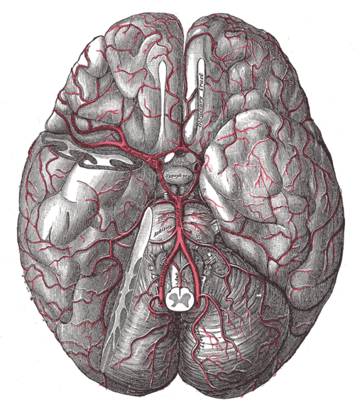
The two vertebral arteries run up the front of the medulla oblongata and join at the pontomedullary junction to form the basilar artery, which ascends in the groove on the front of the pons and divides into the two posterior cerebral arteries at the upper border of the pons/midbrain.
- Branches
- Vertebral: anterior spinal artery (joins its partner — medial medulla, anterior cord), posterior spinal arteries, PICA (lateral medulla, inferior cerebellum)
- Basilar: AICA (lateral lower pons, anterior-inferior cerebellum; often gives the labyrinthine artery to the inner ear), pontine paramedian and circumferential branches, superior cerebellar artery (upper pons, superior cerebellum)
- Posterior cerebral arteries (terminal branches; see cerebral arteries (ACA, MCA, PCA))
- Supplies
Medulla oblongata, pons, midbrain, cerebellum, inner ear, thalamus, occipital and inferior temporal lobes, upper spinal cord.
- Anastomoses
Joins the anterior circulation through the posterior communicating arteries (see circle of Willis).
- Termination
Bifurcates into the posterior cerebral arteries at the midbrain.
- Relations
CN III emerges between the PCA and SCA; CN VI runs up alongside the basilar artery.
Branches
Structures supplied
Generated automatically from links in other entries.
Swipe sideways to see the whole diagram →


Clinical correlations
Posterior circulation stroke PANCE
- Mechanism / lesion
Embolism, atherosclerosis of the vertebral/basilar arteries, vertebral artery dissection (neck trauma or manipulation).
- Classic presentation
"5 Ds": dizziness/vertigo, diplopia, dysarthria, dysphagia, dystaxia (ataxia), plus nausea, nystagmus and crossed signs (ipsilateral cranial nerve + contralateral limb deficits). PICA → lateral medullary (Wallenberg); AICA → lateral pontine syndrome (ipsilateral facial palsy, hearing loss, vertigo, ataxia, face sensory loss, contralateral body pain/temp loss); SCA → ipsilateral ataxia.
Basilar artery occlusion PANCE
- Mechanism / lesion
Thrombosis or embolism — high mortality without reperfusion.
- Classic presentation
Fluctuating or sudden coma, quadriparesis, pinpoint pupils, bilateral signs, or locked-in syndrome; "top of the basilar" syndrome — visual loss, eye movement abnormalities, altered consciousness. Emergency CTA and thrombectomy.
Subclavian steal syndrome PANCE
- Mechanism / lesion
Stenosis of the proximal subclavian artery (before the vertebral origin, usually left) → blood flows retrograde down the ipsilateral vertebral artery to supply the arm during exercise.
- Classic presentation
Arm claudication and vertebrobasilar symptoms (dizziness, syncope) with arm exercise; arm BP difference > 15–20 mm Hg and a weaker radial pulse on the affected side. Duplex shows reversed vertebral flow.
High-yield PANCE pearls
- Vertigo + diplopia + dysarthria + ataxia + crossed signs → posterior circulation stroke → MRI (CT misses it).
- Coma or locked-in state + pinpoint pupils → basilar occlusion → CTA + thrombectomy.
- Dizziness when using one arm + unequal arm BPs → subclavian steal.
- Vertigo + sudden unilateral hearing loss → AICA (labyrinthine artery) stroke.
- Neck pain + posterior circulation stroke in a young patient → vertebral dissection.
Imaging anatomy
- CT
Noncontrast CT has poor sensitivity in the posterior fossa; CTA for vertebral/basilar occlusion and dissection.
- MRI
MRI with DWI is the test of choice for posterior circulation infarcts; MRA and fat-saturated T1 for dissection.
- Ultrasound
Duplex of the vertebral arteries shows reversed flow in subclavian steal.
Physical exam
- Strength / function testing
Eye movements and nystagmus, HINTS exam, facial sensation and strength, hearing, palate and tongue, limb ataxia and gait, crossed sensory findings; blood pressure in both arms and bilateral radial pulses.
Self-quiz
A 66-year-old has dizziness and near-fainting whenever he chops wood with his left arm. BP is 150/85 in the right arm and 118/70 in the left. What is the most likely diagnosis?
Correct answer: B
Proximal left subclavian stenosis makes the arm "steal" blood by reversing flow down the left vertebral artery during exercise, causing posterior circulation symptoms and a large inter-arm BP difference.
Show answer
B
Proximal left subclavian stenosis makes the arm "steal" blood by reversing flow down the left vertebral artery during exercise, causing posterior circulation symptoms and a large inter-arm BP difference.
A 58-year-old has sudden vertigo, vomiting, right-sided hearing loss, right facial weakness and right limb ataxia. Which artery is most likely occluded?
Correct answer: B
AICA supplies the lateral lower pons (facial nucleus, vestibular/cochlear nuclei) and usually the labyrinthine artery — vertigo with hearing loss and facial palsy suggests the lateral pontine (AICA) syndrome.
Show answer
B
AICA supplies the lateral lower pons (facial nucleus, vestibular/cochlear nuclei) and usually the labyrinthine artery — vertigo with hearing loss and facial palsy suggests the lateral pontine (AICA) syndrome.
A 70-year-old is found comatose with quadriparesis and pinpoint pupils. CT shows no hemorrhage. What is the most important next step?
Correct answer: A
Sudden coma, quadriparesis and pinpoint pupils suggest basilar artery occlusion, which has high mortality without reperfusion. It needs emergency CTA and thrombectomy.
Show answer
A
Sudden coma, quadriparesis and pinpoint pupils suggest basilar artery occlusion, which has high mortality without reperfusion. It needs emergency CTA and thrombectomy.
The posterior cerebral arteries are the terminal branches of which artery?
Correct answer: E
The basilar artery divides into the two posterior cerebral arteries at the upper pons/midbrain. The PComm arteries connect them to the internal carotids.
Show answer
E
The basilar artery divides into the two posterior cerebral arteries at the upper pons/midbrain. The PComm arteries connect them to the internal carotids.
Which imaging test best detects an acute posterior circulation (brainstem or cerebellar) stroke?
Correct answer: C
CT often misses posterior fossa infarcts because of bone artifact. MRI (diffusion-weighted) is the test of choice when a posterior circulation stroke is suspected.
Show answer
C
CT often misses posterior fossa infarcts because of bone artifact. MRI (diffusion-weighted) is the test of choice when a posterior circulation stroke is suspected.