Thoracic vertebrae
Twelve vertebrae that articulate with the ribs. They are the common site of osteoporotic compression fractures, metastases and spinal tuberculosis.
Core anatomy
- Location
Posterior thorax, T1–T12.
- Classification
Irregular bones.
- Key features & landmarks
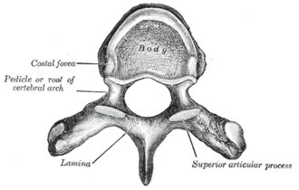
- Costal facets on the bodies (for the heads of the ribs) and on the transverse processes of T1–T10 (for the rib tubercles)
- Heart-shaped bodies; small circular vertebral foramen
- Long spinous processes sloping inferiorly (overlapping the vertebra below in the mid-thoracic region)
- Articular facets oriented roughly in the coronal plane, favoring rotation; motion is limited by the rib cage
- Relations
Articulate with the ribs at costovertebral and costotransverse joints. The descending aorta lies to the left of the bodies and the azygos vein and thoracic duct to the right; the sympathetic trunks lie on the rib heads.
- Notes
The thoracolumbar junction (T11–L2) is a transition from the stiff, rib-supported thoracic spine to the mobile lumbar spine and is the most common site of traumatic fractures.

Clinical correlations
Osteoporotic vertebral compression fracture PANCE
- Mechanism / lesion
Fracture of a weakened vertebral body with minimal trauma (bending, lifting, coughing), often in the mid-thoracic or thoracolumbar region, in older adults, especially postmenopausal women and those on glucocorticoids.
- Classic presentation
Sudden back pain with point tenderness, or silent height loss and progressive kyphosis. A fragility vertebral fracture diagnoses osteoporosis and should prompt DEXA and treatment.
Spinal metastases PANCE
- Mechanism / lesion
Hematogenous spread to vertebral bodies (breast, lung, prostate, kidney, thyroid; multiple myeloma). The thoracic spine is most often involved.
- Classic presentation
Progressive back pain, worse at night or supine, with weight loss or a cancer history. Malignant spinal cord compression (weakness, sensory level, bladder dysfunction) is an emergency: high-dose steroids, MRI of the whole spine, and radiotherapy or surgery.
Spinal tuberculosis (Pott disease)
- Mechanism / lesion
TB spondylitis, usually of the lower thoracic and upper lumbar spine, destroying adjacent vertebral bodies and the disc.
- Classic presentation
Back pain, systemic symptoms, gibbus deformity (sharp angular kyphosis), possible paraspinal/psoas cold abscess and paraplegia.
High-yield PANCE pearls
- Minimal-trauma vertebral fracture = osteoporosis → DEXA and treatment.
- Back pain worse at night/supine + cancer history → metastases; new weakness or sensory level → emergency MRI (cord compression).
- Thoracic spine has costal facets and long downward spinous processes.
- Pott disease: TB of the lower thoracic spine, gibbus, psoas abscess.
Imaging anatomy
- X-ray
Loss of vertebral body height (wedge compression). Pedicle destruction ("winking owl" sign) suggests metastasis on the AP view.
- CT
Fracture morphology and posterior wall involvement (burst fracture).
- MRI
Acute vs chronic compression fracture (marrow edema on STIR), malignant vs osteoporotic fracture, spinal cord compression, infection.
- Key views & landmarks
- Lateral thoracic/lumbar radiographs
- MRI whole spine for suspected cord compression
- DEXA after fragility fracture
Physical exam
- Surface landmarks
T3 at the medial end of the scapular spine; T7 at the inferior angle of the scapula (arm at side); T12 at the lowest rib attachment.
- Palpation
Percussion tenderness over a spinous process (fracture, infection, malignancy). Measure height and look for kyphosis.
- Strength / function testing
Neurologic exam for a sensory level and UMN signs in the legs (cord compression).
Self-quiz
A 74-year-old woman felt sudden mid-back pain after lifting a laundry basket. A radiograph shows anterior wedging of T8. What is the most appropriate next step for long-term management?
Correct answer: B
A low-trauma vertebral compression fracture establishes osteoporosis clinically. Evaluate with DEXA (and secondary causes) and start treatment to prevent further fractures.
Show answer
B
A low-trauma vertebral compression fracture establishes osteoporosis clinically. Evaluate with DEXA (and secondary causes) and start treatment to prevent further fractures.
A 66-year-old man with prostate cancer has 3 weeks of worsening thoracic back pain, now with bilateral leg weakness and urinary retention. What is the most urgent step?
Correct answer: B
This is malignant spinal cord compression, an oncologic emergency: give high-dose steroids and obtain an urgent MRI of the whole spine, followed by radiotherapy and/or surgery.
Show answer
B
This is malignant spinal cord compression, an oncologic emergency: give high-dose steroids and obtain an urgent MRI of the whole spine, followed by radiotherapy and/or surgery.
Which feature is unique to thoracic vertebrae?
Correct answer: C
Thoracic vertebrae have costal facets on their bodies (and most on their transverse processes) for articulation with the ribs.
Show answer
C
Thoracic vertebrae have costal facets on their bodies (and most on their transverse processes) for articulation with the ribs.
A man from a TB-endemic region has back pain, night sweats and a sharp angular kyphosis of the lower thoracic spine. Imaging shows destruction of adjacent vertebral bodies and the disc with a psoas abscess. What is the diagnosis?
Correct answer: B
Pott disease (TB spondylitis) usually affects the lower thoracic and upper lumbar spine, destroying adjacent bodies and the disc, causing a gibbus deformity, possible paraspinal/psoas cold abscess and paraplegia.
Show answer
B
Pott disease (TB spondylitis) usually affects the lower thoracic and upper lumbar spine, destroying adjacent bodies and the disc, causing a gibbus deformity, possible paraspinal/psoas cold abscess and paraplegia.
Where do traumatic spinal fractures most commonly occur?
Correct answer: D
The thoracolumbar junction (T11–L2), where the stiff, rib-supported thoracic spine meets the mobile lumbar spine, is the most common site of traumatic fractures.
Show answer
D
The thoracolumbar junction (T11–L2), where the stiff, rib-supported thoracic spine meets the mobile lumbar spine, is the most common site of traumatic fractures.