Tricuspid valve
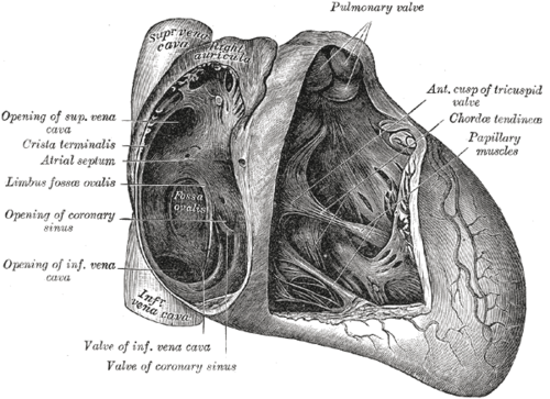
Three-leaflet valve between the right atrium and right ventricle. Tricuspid regurgitation, including from IV drug–associated endocarditis, is its key clinical problem.
Core anatomy
- Location
Between the right atrium and right ventricle, behind the sternum at about the level of the 4th intercostal space. verify
- Parts
- Three leaflets: anterior, posterior and septal
- Chordae tendineae attaching to the anterior, posterior and septal papillary muscles
- Relations
The septal leaflet lies near the membranous septum and the AV conduction tissue.
- Notes
Auscultation area: the left lower sternal border (4th–5th intercostal space).

Clinical correlations
Tricuspid regurgitation PANCE
- Mechanism / lesion
Usually functional (RV dilation from pulmonary hypertension or left heart failure). Primary causes include infective endocarditis in people who inject drugs (commonly Staphylococcus aureus), carcinoid heart disease, Ebstein anomaly, rheumatic disease, and pacemaker/ICD leads.
- Classic presentation
Holosystolic murmur at the left lower sternal border that increases with inspiration (Carvallo sign). Prominent v waves in the JVP, a pulsatile liver, peripheral edema, ascites.
Right-sided endocarditis PANCE
- Mechanism / lesion
Injection drug use; S. aureus is the most common organism.
- Classic presentation
Fever and septic pulmonary emboli (multiple peripheral nodules, often cavitating) with a TR murmur. Left-sided embolic signs are usually absent.
Ebstein anomaly
- Mechanism / lesion
Apical displacement of the septal and posterior leaflets, "atrializing" part of the RV. Associated with maternal lithium exposure.
- Classic presentation
Cyanosis, TR, right heart failure and arrhythmias (associated with WPW).
High-yield PANCE pearls
- TR: holosystolic murmur at the left lower sternal border, louder on inspiration (Carvallo sign); giant v waves in the JVP.
- Injection drug use + fever + murmur → tricuspid endocarditis (S. aureus) with septic pulmonary emboli.
- Ebstein anomaly ↔ lithium exposure in pregnancy.
- Right-sided murmurs increase with inspiration (↑ venous return).
Imaging anatomy
- X-ray
Right atrial enlargement (prominent right heart border). Septic emboli in endocarditis show as multiple peripheral nodules.
- CT
Septic pulmonary emboli (peripheral, cavitating nodules, feeding vessel sign).
- Ultrasound
Echo shows leaflet morphology, vegetations, regurgitant jet severity, RV size and function, and estimates pulmonary artery systolic pressure from the TR jet velocity. Ebstein anomaly shows apical displacement of the septal leaflet.
- Key views & landmarks
- Apical 4-chamber and RV-focused views; subcostal (IVC size)
Physical exam
- Surface landmarks
Tricuspid area: left lower sternal border, 4th–5th intercostal space.
- Palpation
Parasternal (RV) heave. A pulsatile, enlarged liver in severe TR.
- Strength / function testing
Listen at the left lower sternal border during inspiration and expiration: right-sided murmurs intensify with inspiration. Inspect the JVP for large v (cv) waves.
Self-quiz
A 28-year-old man who injects heroin has fever and a holosystolic murmur at the left lower sternal border that increases with inspiration. Chest CT shows multiple peripheral cavitating nodules. What is the most likely organism?
Correct answer: B
Tricuspid valve endocarditis in people who inject drugs is most often caused by S. aureus. The cavitating nodules are septic pulmonary emboli.
Show answer
B
Tricuspid valve endocarditis in people who inject drugs is most often caused by S. aureus. The cavitating nodules are septic pulmonary emboli.
A holosystolic murmur that gets louder during inspiration is most consistent with which lesion?
Correct answer: C
Inspiration increases venous return to the right heart, intensifying right-sided murmurs (Carvallo sign in TR). MR doesn't change or softens with inspiration.
Show answer
C
Inspiration increases venous return to the right heart, intensifying right-sided murmurs (Carvallo sign in TR). MR doesn't change or softens with inspiration.
A newborn whose mother took lithium during pregnancy has cyanosis and an enlarged right atrium. Which valve abnormality is expected?
Correct answer: B
Ebstein anomaly, apical displacement of the septal and posterior tricuspid leaflets, is associated with prenatal lithium exposure.
Show answer
B
Ebstein anomaly, apical displacement of the septal and posterior tricuspid leaflets, is associated with prenatal lithium exposure.
What are the three leaflets of the tricuspid valve?
Correct answer: B
The tricuspid valve has anterior, posterior and septal leaflets. The septal leaflet lies near the membranous septum and AV conduction tissue. The aortic valve has right, left and posterior (non-coronary) cusps.
Show answer
B
The tricuspid valve has anterior, posterior and septal leaflets. The septal leaflet lies near the membranous septum and AV conduction tissue. The aortic valve has right, left and posterior (non-coronary) cusps.
Which jugular venous finding is typical of significant tricuspid regurgitation?
Correct answer: E
Regurgitant flow into the right atrium during systole produces prominent (giant) v waves in the JVP, often with a pulsatile liver. Cannon a waves occur with AV dissociation.
Show answer
E
Regurgitant flow into the right atrium during systole produces prominent (giant) v waves in the JVP, often with a pulsatile liver. Cannon a waves occur with AV dissociation.