Aortic valve
Three-cusped semilunar valve between the left ventricle and the aorta. Aortic stenosis and regurgitation are among the most tested murmurs.
Core anatomy
- Location
At the outflow of the left ventricle, posterior to the left side of the sternum at about the level of the 3rd intercostal space. verify
- Parts
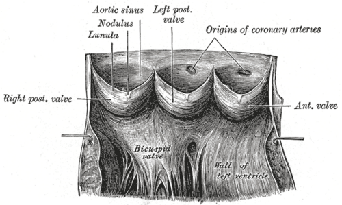
- Three semilunar cusps: right, left and posterior (non-coronary)
- Aortic sinuses (of Valsalva): dilations above each cusp. The right coronary artery arises from the right sinus and the left coronary artery from the left sinus
- Normally tricuspid. A bicuspid aortic valve is the most common congenital heart anomaly
- Relations
Continuous with the anterior leaflet of the mitral valve (aortomitral continuity). The membranous septum and His bundle lie just below.
- Arterial supply
Avascular cusps (nourished by diffusion). The coronary ostia sit in the sinuses just above the cusps.
- Notes
Auscultation area: the 2nd right intercostal space at the right sternal border. Aortic regurgitation is often best heard at the left sternal border (Erb's point, 3rd left intercostal space), with the patient leaning forward in held expiration.
Mentioned in
Generated automatically from links in other entries.

Clinical correlations
Aortic stenosis PANCE
- Mechanism / lesion
Age-related calcific degeneration (older adults), a bicuspid valve (earlier presentation, often 40s–60s), or rheumatic disease (usually with mitral involvement).
- Classic presentation
Symptoms: syncope, angina and dyspnea (heart failure), classically exertional. Crescendo–decrescendo systolic ejection murmur at the right 2nd intercostal space radiating to the carotids. Pulsus parvus et tardus (weak, delayed carotid upstroke). A soft or absent A2 and a late-peaking murmur indicate severe stenosis. Symptomatic severe AS is an indication for valve replacement.
Aortic regurgitation PANCE
- Mechanism / lesion
Leaflet disease (bicuspid valve, endocarditis, rheumatic) or aortic root dilation (hypertension, Marfan syndrome, aortic dissection, syphilitic aortitis, ankylosing spondylitis).
- Classic presentation
Early diastolic, blowing, decrescendo murmur at the left sternal border, best with the patient sitting forward in held expiration. Wide pulse pressure with bounding pulses ("water hammer"/Corrigan pulse, de Musset head bobbing, Quincke capillary pulsations). An Austin Flint mid-diastolic rumble may be heard at the apex. Acute AR (dissection, endocarditis) causes sudden pulmonary edema and shock.
Infective endocarditis PANCE
- Mechanism / lesion
Bacterial colonization of the valve, more likely with abnormal or prosthetic valves.
- Classic presentation
Fever, a new regurgitant murmur, embolic phenomena (splinter hemorrhages, Janeway lesions), and immunologic signs (Osler nodes, Roth spots). Diagnosed with blood cultures and echocardiography (modified Duke criteria).
High-yield PANCE pearls
- AS: syncope, angina, dyspnea; crescendo–decrescendo systolic murmur at the right 2nd ICS → carotids; pulsus parvus et tardus.
- AR: early diastolic decrescendo murmur at the left sternal border; wide pulse pressure, bounding pulses.
- Bicuspid aortic valve: most common congenital heart defect; associated with coarctation and Turner syndrome; aortic root dilation.
- Handgrip increases AR (↑ afterload) and decreases AS; Valsalva and standing decrease the AS murmur (↓ preload).
Imaging anatomy
- X-ray
Post-stenotic dilation of the ascending aorta; valve calcification (better on lateral). LV enlargement in chronic AR.
- CT
Aortic valve calcium scoring (helps grade AS); CT for sizing before transcatheter aortic valve replacement (TAVR); aortic root dimensions.
- MRI
Quantifies regurgitant volume and aortic dimensions.
- Ultrasound
Echocardiography is the key test: valve morphology (bicuspid vs tricuspid), severity of AS (peak velocity, mean gradient, valve area) and AR (jet width, vena contracta), LV size and function, and aortic root size. TEE for endocarditis (vegetations, abscess) and better morphology.
- Key views & landmarks
- Parasternal long axis (LVOT, root) and short axis (cusp number: the 'Mercedes-Benz' sign of a normal trileaflet valve)
- Apical 5-chamber with continuous-wave Doppler for gradients
Physical exam
- Surface landmarks
Aortic area: right 2nd intercostal space at the sternal border.
- Palpation
Carotid pulse (slow-rising and weak in AS; bounding and collapsing in AR). A systolic thrill may be palpable at the base in severe AS.
- Strength / function testing
Auscultate with the diaphragm at the aortic area and along the left sternal border. For AR, have the patient sit up, lean forward and exhale fully. Measure BP for pulse pressure. Dynamic maneuvers: handgrip (increases afterload) intensifies regurgitant murmurs; Valsalva/standing (decreases preload) softens the AS murmur.
Self-quiz
A 78-year-old man has exertional chest pain and two episodes of syncope. There is a harsh crescendo–decrescendo systolic murmur at the right upper sternal border radiating to both carotids, and the carotid upstroke is weak and delayed. What is the most likely diagnosis?
Correct answer: C
Aortic stenosis: exertional syncope and angina, an ejection murmur at the aortic area radiating to the carotids, and pulsus parvus et tardus. In an older patient, calcific degeneration is the usual cause.
Show answer
C
Aortic stenosis: exertional syncope and angina, an ejection murmur at the aortic area radiating to the carotids, and pulsus parvus et tardus. In an older patient, calcific degeneration is the usual cause.
A 25-year-old tall man with long fingers and lens dislocation has a blowing early diastolic murmur at the left sternal border and a BP of 150/45 mmHg. What is the mechanism of his murmur?
Correct answer: B
Marfan syndrome causes aortic root dilation, which pulls the cusps apart and causes aortic regurgitation (early diastolic decrescendo murmur, wide pulse pressure). Mitral valve prolapse is also common in Marfan but gives a systolic click.
Show answer
B
Marfan syndrome causes aortic root dilation, which pulls the cusps apart and causes aortic regurgitation (early diastolic decrescendo murmur, wide pulse pressure). Mitral valve prolapse is also common in Marfan but gives a systolic click.
Where is the aortic valve area for auscultation?
Correct answer: B
The aortic area is the right 2nd intercostal space at the sternal border. The pulmonic area is the left 2nd, the tricuspid area the left lower sternal border, and the mitral area the apex.
Show answer
B
The aortic area is the right 2nd intercostal space at the sternal border. The pulmonic area is the left 2nd, the tricuspid area the left lower sternal border, and the mitral area the apex.
A girl with Turner syndrome is most likely to have which valve abnormality?
Correct answer: C
A bicuspid aortic valve, the most common congenital heart anomaly, is associated with Turner syndrome and coarctation of the aorta, and with aortic root dilation. It can cause aortic stenosis earlier in life.
Show answer
C
A bicuspid aortic valve, the most common congenital heart anomaly, is associated with Turner syndrome and coarctation of the aorta, and with aortic root dilation. It can cause aortic stenosis earlier in life.
How does sustained handgrip affect the murmurs of aortic stenosis and aortic regurgitation?
Correct answer: D
Handgrip raises afterload, which increases regurgitant flow (AR louder) and reduces the gradient across the aortic valve (AS softer). Valsalva and standing reduce preload and soften the AS murmur.
Show answer
D
Handgrip raises afterload, which increases regurgitant flow (AR louder) and reduces the gradient across the aortic valve (AS softer). Valsalva and standing reduce preload and soften the AS murmur.