Corticospinal tract
Main voluntary motor pathway from the motor cortex to the spinal cord. It crosses in the lower medulla; damage causes upper motor neuron signs — weakness, spasticity, hyperreflexia and an extensor plantar response.
Core anatomy
- Origin
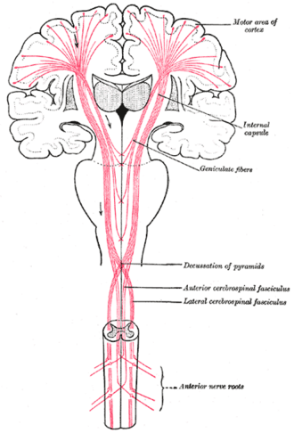
Upper motor neurons in the primary motor cortex (precentral gyrus, including the giant Betz cells of layer V), premotor and supplementary motor areas, and parietal cortex.
- Course
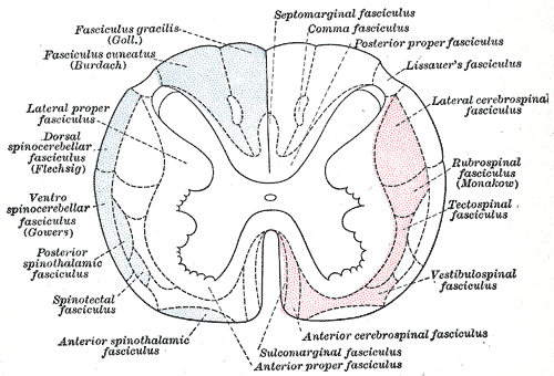
Corona radiata → posterior limb of the internal capsule → middle of the cerebral peduncle (midbrain) → scattered bundles in the basilar pons → pyramids of the medulla oblongata → 85–90% cross in the pyramidal decussation and descend in the lateral corticospinal tract of the contralateral cord → synapse on lower motor neurons in the ventral horn (directly or via interneurons). The uncrossed 10–15% descend in the anterior corticospinal tract (axial muscles) and mostly cross at segmental levels.
- Motor
Voluntary skilled movement, especially of the distal limbs (fine finger movements); lower motor neurons carry the signal to muscle via the spinal nerves.
- Relations
Corticobulbar fibers accompany it to cranial nerve motor nuclei (most receive bilateral input, except the lower face and — partly — CN XII, which are mainly contralateral).
- Notes
A lesion above the decussation (cortex, capsule, brainstem) causes contralateral weakness; a spinal cord lesion causes ipsilateral weakness below it.
Mentioned in
Generated automatically from links in other entries.
Swipe sideways to see the whole diagram →


Clinical correlations
Upper vs lower motor neuron lesions PANCE
- Mechanism / lesion
UMN — damage anywhere along the corticospinal tract (stroke, MS, cord compression, B12 deficiency). LMN — ventral horn cell, root, plexus or peripheral nerve.
- Classic presentation
UMN: weakness in a pyramidal pattern (arm extensors, leg flexors), spasticity (velocity-dependent, "clasp-knife"), hyperreflexia, clonus, Babinski sign (extensor plantar), Hoffmann sign, little atrophy. Acutely (spinal shock) tone and reflexes may be reduced. LMN: weakness, atrophy, fasciculations, hypotonia, hyporeflexia, flexor plantar.
Amyotrophic lateral sclerosis (ALS) PANCE
- Mechanism / lesion
Degeneration of both upper and lower motor neurons (corticospinal tracts and ventral horn/brainstem motor nuclei); sensation spared.
- Classic presentation
Mixed UMN and LMN signs in the same limb — weakness, atrophy, fasciculations with hyperreflexia and Babinski; bulbar dysarthria and dysphagia; no sensory loss. EMG; riluzole; respiratory failure is the usual cause of death.
Cervical spondylotic myelopathy PANCE
- Mechanism / lesion
Degenerative narrowing of the cervical canal compressing the cord (older adults).
- Classic presentation
Clumsy hands, gait imbalance, UMN signs in the legs (and arms below the level), positive Hoffmann sign, bladder urgency; neck pain may be minimal. MRI cervical spine; surgical decompression if progressive.
High-yield PANCE pearls
- UMN: spasticity, hyperreflexia, clonus, Babinski; LMN: atrophy, fasciculations, hyporeflexia.
- UMN + LMN signs, no sensory loss → ALS.
- Crosses at the pyramidal decussation (lower medulla) → brain lesions give contralateral weakness, cord lesions ipsilateral.
- Older adult with clumsy hands, unsteady gait, Hoffmann sign → cervical myelopathy → MRI.
Imaging anatomy
- CT
Noncontrast CT head in acute weakness (stroke protocol).
- MRI
MRI brain or spine at the suspected level — stroke (DWI), demyelination, cord compression; EMG/nerve conduction studies to confirm LMN disease (ALS).
Physical exam
- Strength / function testing
Pronator drift (arms outstretched, palms up, eyes closed — the weak arm pronates and drifts down); strength grading (0–5); tone (spasticity); reflexes; plantar response; clonus at the ankle; Hoffmann sign; gait (circumduction).
Babinski sign (plantar response)
- Technique
Stroke the lateral sole firmly from the heel toward the little toe and across the ball of the foot.
- Positive test
Extension (dorsiflexion) of the great toe, with fanning of the other toes.
- Significance
Upper motor neuron lesion (corticospinal tract). Normal in infants under about 1–2 years.
Hoffmann sign
- Technique
Flick the nail of the relaxed middle finger downward.
- Positive test
Flexion of the thumb and index finger.
- Significance
Suggests an upper motor neuron lesion affecting the arms, e.g. cervical myelopathy (can occur in some normal people).
Self-quiz
A 58-year-old has progressive weakness of the right hand with wasting and fasciculations, brisk reflexes in all limbs, and bilateral Babinski signs. Sensation is normal. What is the most likely diagnosis?
Correct answer: B
Combined UMN (hyperreflexia, Babinski) and LMN (atrophy, fasciculations) signs without sensory loss are the hallmark of ALS.
Show answer
B
Combined UMN (hyperreflexia, Babinski) and LMN (atrophy, fasciculations) signs without sensory loss are the hallmark of ALS.
Which finding indicates an upper motor neuron lesion?
Correct answer: C
An extensor plantar response (Babinski sign) indicates corticospinal tract (UMN) dysfunction. The other findings suggest a lower motor neuron lesion.
Show answer
C
An extensor plantar response (Babinski sign) indicates corticospinal tract (UMN) dysfunction. The other findings suggest a lower motor neuron lesion.
Where do most corticospinal fibers cross the midline?
Correct answer: B
About 85–90% of corticospinal fibers cross in the pyramidal decussation at the medulla–cord junction. Lesions above it cause contralateral weakness; cord lesions cause ipsilateral weakness.
Show answer
B
About 85–90% of corticospinal fibers cross in the pyramidal decussation at the medulla–cord junction. Lesions above it cause contralateral weakness; cord lesions cause ipsilateral weakness.
Immediately after an acute complete spinal cord injury, what are tone and reflexes like below the lesion?
Correct answer: D
In spinal shock, tone and reflexes are initially reduced or absent below the lesion. Upper motor neuron signs (spasticity, hyperreflexia, Babinski) develop later.
Show answer
D
In spinal shock, tone and reflexes are initially reduced or absent below the lesion. Upper motor neuron signs (spasticity, hyperreflexia, Babinski) develop later.
A cortical stroke causes weakness of the opposite lower face but spares the forehead. Why?
Correct answer: C
Most cranial motor nuclei receive bilateral corticobulbar input, but the part of the facial nucleus for the lower face receives mainly contralateral input, so an upper motor neuron lesion weakens only the opposite lower face.
Show answer
C
Most cranial motor nuclei receive bilateral corticobulbar input, but the part of the facial nucleus for the lower face receives mainly contralateral input, so an upper motor neuron lesion weakens only the opposite lower face.