Popliteal fossa
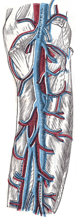
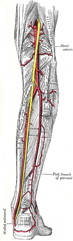
Diamond-shaped space behind the knee containing the popliteal vessels, the tibial and common fibular nerves and lymph nodes. Baker cysts, popliteal aneurysms and knee-dislocation vascular injury arise here.
Core anatomy
- Location
Posterior to the knee joint.
- Boundaries
- superolateral
- Biceps femoris (hamstrings)
- superomedial
- Semimembranosus and semitendinosus
- inferolateral
- Lateral head of gastrocnemius (and plantaris)
- inferomedial
- Medial head of gastrocnemius
- roof
- Skin, superficial fascia (with the small saphenous vein) and the popliteal fascia
- floor
- Popliteal surface of the femur, the posterior knee capsule (oblique popliteal ligament) and popliteus
- Contents
- Superficial to deep: Tibial nerve → popliteal vein → popliteal artery (deepest)
- Common fibular nerve along the lateral border (medial edge of biceps femoris)
- Termination of the small saphenous vein into the popliteal vein
- Popliteal lymph nodes
- Fat and bursae (e.g. semimembranosus–gastrocnemius bursa → Baker cyst)
- Communications
Above with the adductor canal (via the adductor hiatus); below with the posterior compartment of the leg (under the soleal arch).
Mentioned in
- Adductor muscles of the thigh
- Common fibular nerve
- Hamstrings
- Knee joint
- Popliteal artery
- Sciatic nerve
- Tibial nerve
Generated automatically from links in other entries.


Clinical correlations
Popliteal (Baker) cyst PANCE
- Mechanism / lesion
Distension of the gastrocnemius–semimembranosus bursa by knee effusions (OA, RA, meniscal tears).
- Classic presentation
Posteromedial swelling behind the knee, more prominent with the knee extended; rupture mimics DVT (calf pain, swelling) — ultrasound distinguishes them.
Popliteal artery aneurysm and injury PANCE
- Mechanism / lesion
See popliteal artery — atherosclerotic aneurysm or tethered-vessel injury in knee dislocation.
- Classic presentation
Pulsatile mass or distal ischemia; check the ABI after knee dislocation.
Popliteal fossa masses
- Mechanism / lesion
Cysts, aneurysms, lymphadenopathy (foot/heel infections), nerve sheath tumors, sarcomas.
- Classic presentation
A non-pulsatile firm mass that isn't a simple cyst on ultrasound needs further imaging (MRI).
High-yield PANCE pearls
- Popliteal fossa superficial → deep: nerve, vein, artery.
- Posteromedial swelling behind the arthritic knee → Baker cyst; rupture mimics DVT.
- Pulsatile popliteal mass → aneurysm (check the aorta and other knee).
Imaging anatomy
- MRI
Defines masses, cysts and their communication with the joint.
- Ultrasound
First-line for popliteal masses (cyst vs aneurysm vs DVT).
- Key views & landmarks
- Ultrasound of the popliteal fossa
Physical exam
- Surface landmarks
With the knee slightly flexed, the hamstring tendons form the upper borders (biceps laterally; semitendinosus medially) and the gastrocnemius heads the lower.
- Palpation
Palpate with the knee flexed about 30° (relaxed fascia) for the popliteal pulse and masses; a Baker cyst is firmer in extension and softer in flexion (Foucher sign).
Self-quiz
Which structure is the deepest in the popliteal fossa?
Correct answer: C
From superficial to deep: tibial nerve, popliteal vein, popliteal artery. The artery lies against the femur and knee capsule, which is why it is injured in supracondylar fractures and knee dislocations.
Show answer
C
From superficial to deep: tibial nerve, popliteal vein, popliteal artery. The artery lies against the femur and knee capsule, which is why it is injured in supracondylar fractures and knee dislocations.
Which muscle forms the superolateral border of the popliteal fossa?
Correct answer: B
Biceps femoris forms the superolateral border; semimembranosus and semitendinosus the superomedial; the two heads of gastrocnemius the inferior borders.
Show answer
B
Biceps femoris forms the superolateral border; semimembranosus and semitendinosus the superomedial; the two heads of gastrocnemius the inferior borders.
Which muscles form the superomedial border of the popliteal fossa?
Correct answer: B
The popliteal fossa is bounded superomedially by semimembranosus and semitendinosus, superolaterally by biceps femoris, and inferiorly by the two heads of gastrocnemius (with plantaris laterally). Popliteus forms part of the floor.
Show answer
B
The popliteal fossa is bounded superomedially by semimembranosus and semitendinosus, superolaterally by biceps femoris, and inferiorly by the two heads of gastrocnemius (with plantaris laterally). Popliteus forms part of the floor.
Which superficial vein ends in the popliteal fossa by joining the popliteal vein?
Correct answer: D
The small saphenous vein runs up the back of the calf and pierces the popliteal fascia to join the popliteal vein. The great saphenous vein drains into the femoral vein at the saphenous opening.
Show answer
D
The small saphenous vein runs up the back of the calf and pierces the popliteal fascia to join the popliteal vein. The great saphenous vein drains into the femoral vein at the saphenous opening.
A 64-year-old with knee osteoarthritis has a soft posteromedial swelling behind the knee that is more prominent with the knee extended. Which bursa is distended?
Correct answer: E
A popliteal (Baker) cyst is a distended gastrocnemius–semimembranosus bursa, filled by knee effusions from OA, RA or meniscal tears. Its rupture causes calf pain and swelling that mimics DVT.
Show answer
E
A popliteal (Baker) cyst is a distended gastrocnemius–semimembranosus bursa, filled by knee effusions from OA, RA or meniscal tears. Its rupture causes calf pain and swelling that mimics DVT.