Cerebral arteries (ACA, MCA, PCA)
The three paired arteries that supply the cerebral hemispheres. Each territory gives a recognizable stroke syndrome; the MCA is the most commonly affected.
Core anatomy
- Origin
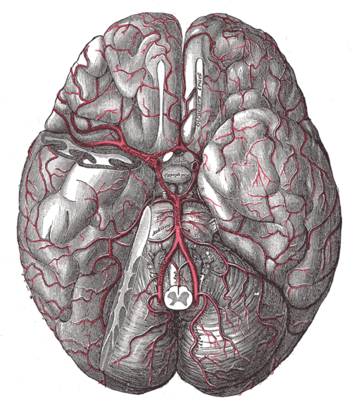
ACA and MCA are the terminal branches of the internal carotid artery (common and internal carotid arteries); the PCAs are the terminal branches of the basilar artery (vertebrobasilar system); all are linked by the circle of Willis.
- Course
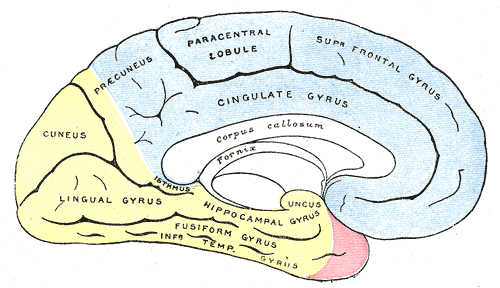
ACA runs forward above the optic chiasm into the longitudinal fissure and arches back over the corpus callosum along the medial surface. MCA runs laterally in the lateral (Sylvian) fissure, giving the lenticulostriate arteries, then superior and inferior divisions over the lateral surface. PCA curves around the midbrain to the occipital lobe and inferior temporal lobe.
- Branches
- ACA: recurrent artery of Heubner, orbitofrontal, callosomarginal, pericallosal
- MCA: lenticulostriate arteries (basal ganglia, internal capsule), superior division (frontal — Broca, motor face/arm), inferior division (temporal/parietal — Wernicke, optic radiations)
- PCA: thalamoperforating and thalamogeniculate branches, posterior choroidal, temporal and calcarine branches
- Supplies
ACA: medial frontal and parietal lobes — motor/sensory cortex for the leg and foot, bladder control, corpus callosum. MCA: most of the lateral hemisphere — face and arm motor/sensory cortex, language areas (dominant), spatial attention (nondominant), optic radiations; deep structures via lenticulostriates. PCA: occipital lobe (visual cortex), inferior temporal lobe (hippocampus), thalamus, midbrain.
- Anastomoses
Leptomeningeal (pial) collaterals link the ends of the territories — the watershed zones between ACA–MCA and MCA–PCA.
Structures supplied
Mentioned in
Generated automatically from links in other entries.
Swipe sideways to see the whole diagram →
Swipe sideways to see the whole diagram →


Clinical correlations
MCA stroke PANCE
- Mechanism / lesion
Embolism (atrial fibrillation, carotid plaque) or thrombosis — the most common territory.
- Classic presentation
Contralateral weakness and sensory loss of the face and arm more than the leg, contralateral homonymous hemianopia, gaze deviation toward the lesion. Dominant (left): aphasia. Nondominant (right): hemineglect, anosognosia.
ACA and PCA strokes PANCE
- Mechanism / lesion
Embolic or thrombotic occlusion.
- Classic presentation
ACA: contralateral leg > arm weakness and sensory loss, urinary incontinence, abulia/personality change. PCA: contralateral homonymous hemianopia with macular sparing, visual agnosia; dominant PCA + splenium → alexia without agraphia; thalamic involvement → sensory loss.
Acute ischemic stroke management PANCE
- Mechanism / lesion
Time-critical reperfusion of the ischemic penumbra.
- Classic presentation
Check glucose, noncontrast CT (exclude hemorrhage), NIHSS. IV thrombolysis (alteplase or tenecteplase) within 4.5 hours of last known well if no contraindications (BP must be < 185/110). Mechanical thrombectomy for large vessel occlusion (ICA, M1) — up to 24 hours in selected patients with CT perfusion/MRI. Then antiplatelet therapy, statin and a search for the source (ECG/telemetry, echocardiogram, carotid imaging).
High-yield PANCE pearls
- Face/arm > leg weakness + aphasia (left) or neglect (right) → MCA.
- Leg > arm weakness + incontinence → ACA.
- Homonymous hemianopia with macular sparing → PCA.
- Eyes deviate toward the lesion in a hemispheric stroke (away from the weak side).
- IV thrombolysis ≤ 4.5 h; thrombectomy for LVO up to 24 h in selected patients.
- Noncontrast CT first — hemorrhage must be excluded before thrombolysis.
Imaging anatomy
- CT
Noncontrast CT (hemorrhage, early ischemic change, hyperdense MCA sign), CT angiography (large vessel occlusion), CT perfusion (core vs penumbra).
- MRI
DWI is the most sensitive test for acute infarction.
- Key views & landmarks
- Stroke code: glucose → noncontrast CT → CTA ± CTP
Physical exam
- Strength / function testing
NIH Stroke Scale (consciousness, gaze, visual fields, facial palsy, arm/leg drift, ataxia, sensation, language, dysarthria, neglect); compare face vs arm vs leg; check for aphasia, neglect and field cuts.
Self-quiz
A 70-year-old with atrial fibrillation suddenly develops right face and arm weakness, right homonymous hemianopia, and difficulty producing and understanding speech. His eyes deviate to the left. Which artery is occluded?
Correct answer: B
Contralateral face/arm weakness, field cut, aphasia (dominant hemisphere) and gaze toward the lesion point to a left MCA occlusion — likely cardioembolic.
Show answer
B
Contralateral face/arm weakness, field cut, aphasia (dominant hemisphere) and gaze toward the lesion point to a left MCA occlusion — likely cardioembolic.
A 64-year-old develops weakness and numbness of the left leg, with only mild left arm weakness, and new urinary incontinence. Which artery is involved?
Correct answer: A
The leg area of the motor and sensory cortex is on the medial surface, supplied by the ACA; a right ACA stroke causes left leg-predominant weakness and often incontinence.
Show answer
A
The leg area of the motor and sensory cortex is on the medial surface, supplied by the ACA; a right ACA stroke causes left leg-predominant weakness and often incontinence.
A 78-year-old has sudden loss of the left visual field in both eyes. Central vision is preserved, and there is no weakness. Which artery is most likely occluded?
Correct answer: A
A left homonymous hemianopia with macular sparing and no weakness localizes to the right occipital lobe — right PCA territory.
Show answer
A
A left homonymous hemianopia with macular sparing and no weakness localizes to the right occipital lobe — right PCA territory.
In an acute hemispheric (cortical) stroke, which way do the eyes usually deviate?
Correct answer: A
A destructive hemispheric lesion knocks out the frontal eye field, so the intact opposite field drives the eyes toward the lesion, away from the hemiparesis.
Show answer
A
A destructive hemispheric lesion knocks out the frontal eye field, so the intact opposite field drives the eyes toward the lesion, away from the hemiparesis.
The lenticulostriate arteries supplying the basal ganglia and internal capsule arise from which artery?
Correct answer: E
The lenticulostriate arteries are branches of the MCA. They are the vessels involved in lacunar infarcts and hypertensive hemorrhage of the putamen and internal capsule.
Show answer
E
The lenticulostriate arteries are branches of the MCA. They are the vessels involved in lacunar infarcts and hypertensive hemorrhage of the putamen and internal capsule.