Colon
Large intestine from the cecum to the sigmoid colon, supplied by the SMA (midgut) and IMA (hindgut). Diverticulitis, colorectal cancer, volvulus and ischemic colitis are the high-yield problems.
Core anatomy
- Location
Frames the small intestine around the abdomen.
- Parts
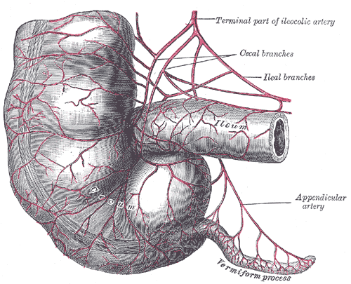
- Cecum (right iliac fossa, with the vermiform appendix) and ileocecal valve
- Ascending colon (retroperitoneal) → right colic (hepatic) flexure
- Transverse colon (intraperitoneal, on the transverse mesocolon) → left colic (splenic) flexure (higher than the hepatic flexure)
- Descending colon (retroperitoneal)
- Sigmoid colon (intraperitoneal, on the sigmoid mesocolon) → rectum at about S3
- Distinguishing features: teniae coli (three longitudinal muscle bands), haustra, omental appendices (fat tags)
- Relations
The ascending and descending colon lie on the posterior abdominal wall (kidneys, quadratus lumborum). The transverse colon is attached to the greater omentum. Paracolic gutters lie lateral to the ascending and descending colon.
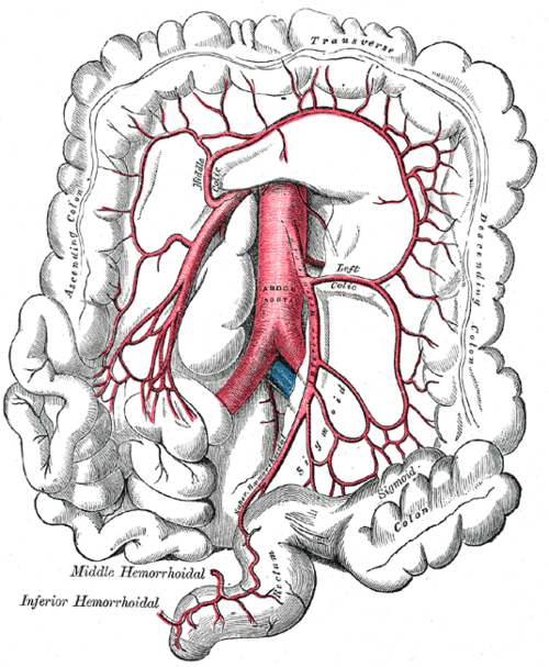
- Arterial supply
Midgut (cecum to about the distal third of the transverse colon): Superior mesenteric artery via the ileocolic, right colic and middle colic arteries. Hindgut (distal third of the transverse colon to the upper rectum): Inferior mesenteric artery via the left colic, sigmoid and superior rectal arteries. The marginal artery (of Drummond) runs along the mesenteric border, connecting them. Watershed areas at the splenic flexure (Griffiths point) and the rectosigmoid junction (Sudeck point) are vulnerable to low-flow ischemia.
- Venous drainage
Superior and inferior mesenteric veins → hepatic portal vein (the IMV usually joins the splenic vein).
- Lymphatic drainage
Epicolic → paracolic → intermediate (along the colic arteries) → superior and inferior mesenteric nodes.
- Innervation
Vagus to about the distal transverse colon; pelvic splanchnic nerves (S2–S4) beyond; sympathetics via the superior and inferior mesenteric plexuses. Visceral pain is periumbilical (midgut) or suprapubic (hindgut).


Clinical correlations
Diverticulosis and diverticulitis PANCE
- Mechanism / lesion
Outpouchings of mucosa and submucosa through weak points where vasa recta penetrate the wall, most often in the sigmoid (in Western populations). Associated with low-fiber diets and age.
- Classic presentation
Diverticulosis: usually asymptomatic; painless lower GI bleeding (the most common cause of significant hematochezia in older adults). Diverticulitis: LLQ pain, fever, leukocytosis, altered bowel habit. Complications: abscess, perforation, fistula (colovesical → pneumaturia), stricture.
Colorectal cancer PANCE
- Mechanism / lesion
Adenoma–carcinoma sequence. Risk factors include age, family history, hereditary syndromes (Lynch, FAP), inflammatory bowel disease, and diet.
- Classic presentation
Right-sided tumors: iron-deficiency anemia, occult bleeding, fatigue, weight loss. Left-sided tumors: obstruction, change in bowel habit, narrowed stool caliber, hematochezia. Iron-deficiency anemia in an older adult (or a postmenopausal woman) warrants colonoscopy. Screening typically starts at age 45 for average-risk adults.
Volvulus PANCE
- Mechanism / lesion
Twisting of a mobile segment on its mesentery. Sigmoid volvulus is most common (older, institutionalized or constipated patients); cecal volvulus occurs in younger patients.
- Classic presentation
Abdominal distension, pain, obstipation. Sigmoid volvulus shows a "coffee bean" sign on X-ray and is often treated first with endoscopic detorsion (if no ischemia), then elective resection.
Ischemic colitis PANCE
- Mechanism / lesion
Low-flow ischemia at the watershed areas (splenic flexure, rectosigmoid), e.g. with hypotension, vascular disease, or after aortic surgery with IMA ligation.
- Classic presentation
Crampy LLQ pain followed within 24 hours by bloody diarrhea, in an older patient. "Thumbprinting" on imaging.
High-yield PANCE pearls
- Diverticulitis: LLQ pain + fever; diagnose with CT; avoid colonoscopy in the acute phase (colonoscopy afterward to exclude cancer).
- Painless hematochezia in an older adult → diverticulosis (or angiodysplasia).
- Right colon cancer → iron-deficiency anemia; left colon cancer → obstruction, change in stool caliber.
- Sigmoid volvulus: coffee bean sign; endoscopic detorsion.
- Watershed areas: splenic flexure and rectosigmoid → ischemic colitis.
- In large bowel obstruction the cecum is most at risk of perforation (widest diameter, Laplace law).
Imaging anatomy
- X-ray
Large bowel obstruction: dilated colon with haustral markings at the periphery (the cecum above roughly 9–12 cm is at risk of perforation). Coffee bean sign in sigmoid volvulus. Free air. verify
- CT
CT with IV contrast is the test of choice for diverticulitis (wall thickening, pericolic fat stranding, abscess) and for obstruction, volvulus (whirl sign), ischemic colitis and cancer staging. CT colonography is a screening alternative.
- Ultrasound
Useful in some centers for diverticulitis; limited by gas.
- Key views & landmarks
- Colonoscopy: gold standard for cancer detection and polyp removal
- Barium enema (historic): apple-core lesion of colon cancer
Physical exam
- Surface landmarks
The cecum lies in the right iliac fossa; the sigmoid in the left iliac fossa (a firm, stool-filled sigmoid may be palpable).
- Palpation
LLQ tenderness (diverticulitis), masses, distension. Digital rectal exam for masses, blood and tone.
- Strength / function testing
Percuss for tympany in distension. Check stool for occult blood when indicated.
Self-quiz
A 67-year-old man has 3 days of LLQ pain, fever of 38.4 °C and leukocytosis. What is the best diagnostic test?
Correct answer: B
Suspected acute diverticulitis is best confirmed with CT, which also detects abscess or perforation. Colonoscopy and barium enema are avoided acutely (perforation risk).
Show answer
B
Suspected acute diverticulitis is best confirmed with CT, which also detects abscess or perforation. Colonoscopy and barium enema are avoided acutely (perforation risk).
A 70-year-old woman has fatigue and iron-deficiency anemia with no visible bleeding. Where is a colorectal cancer most likely to cause this presentation?
Correct answer: D
Right-sided (cecal/ascending) cancers bleed occultly and present with iron-deficiency anemia. Left-sided cancers tend to cause obstruction and change in bowel habits.
Show answer
D
Right-sided (cecal/ascending) cancers bleed occultly and present with iron-deficiency anemia. Left-sided cancers tend to cause obstruction and change in bowel habits.
After an abdominal aortic aneurysm repair with ligation of the IMA, a patient develops bloody diarrhea. Which part of the colon is most at risk?
Correct answer: C
The IMA supplies the distal colon. With it ligated, the splenic flexure and rectosigmoid (sigmoid) watershed regions depend on collaterals and are prone to ischemic colitis.
Show answer
C
The IMA supplies the distal colon. With it ligated, the splenic flexure and rectosigmoid (sigmoid) watershed regions depend on collaterals and are prone to ischemic colitis.
An elderly nursing home resident has massive abdominal distension and obstipation. The radiograph shows a large inverted-U loop resembling a coffee bean pointing toward the right upper quadrant. What is the diagnosis?
Correct answer: B
The coffee bean sign in an older, institutionalized patient is classic for sigmoid volvulus. Treatment starts with endoscopic detorsion if there's no ischemia.
Show answer
B
The coffee bean sign in an older, institutionalized patient is classic for sigmoid volvulus. Treatment starts with endoscopic detorsion if there's no ischemia.
In a large bowel obstruction with a competent ileocecal valve, which part of the colon is most at risk of perforation?
Correct answer: E
The cecum has the widest diameter, so by the law of Laplace its wall tension is highest and it perforates first in a closed-loop large bowel obstruction.
Show answer
E
The cecum has the widest diameter, so by the law of Laplace its wall tension is highest and it perforates first in a closed-loop large bowel obstruction.