Abducens nerve (CN VI)
Supplies the lateral rectus. Its long course along the clivus and through the cavernous sinus makes it the cranial nerve most often affected by raised intracranial pressure, causing horizontal diplopia.
Core anatomy
- Origin
Abducens nucleus in the floor of the fourth ventricle in the caudal pons; facial nerve fibers loop around it, forming the facial colliculus. The nerve emerges at the pontomedullary junction.
- Course
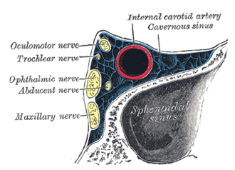
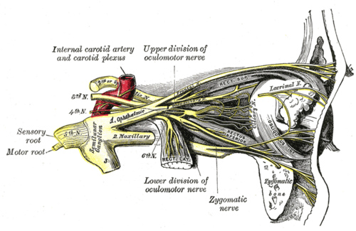
Ascends along the clivus, crosses the petrous apex under the petrosphenoid ligament (Dorello canal), runs inside the cavernous sinus beside the internal carotid artery (the only nerve within the sinus itself — the others are in its wall; see dural venous sinuses), and enters the orbit through the superior orbital fissure within the common tendinous ring.
- Motor
Lateral rectus only — abduction of the eye (see extraocular muscles). Mnemonic LR6 SO4.
- Relations
The abducens nucleus also contains interneurons that project through the medial longitudinal fasciculus (MLF) to the contralateral medial rectus subnucleus, coordinating horizontal gaze.
Muscles innervated
- Extraocular muscles — LR6 SO4, rest 3: lateral rectus — abducens nerve (CN VI); superior oblique — trochlear nerve (CN IV); superior, inferior and medial recti and inferior oblique — oculomotor nerve (CN III, which also supplies levator palpebrae superioris).
Found in
Mentioned in
Generated automatically from links in other entries.
Swipe sideways to see the whole diagram →


Clinical correlations
Sixth nerve palsy PANCE
- Mechanism / lesion
Raised intracranial pressure (a "false localizing sign" — the nerve is stretched along the clivus; e.g. idiopathic intracranial hypertension, tumors), microvascular ischemia (diabetes, hypertension), cavernous sinus lesions (thrombosis, carotid aneurysm, carotid–cavernous fistula), trauma, and after lumbar puncture.
- Classic presentation
Horizontal diplopia worse at distance and on looking toward the affected side; the eye is turned in (esotropia) and cannot abduct; patients may turn the head toward the affected side.
Abducens nucleus and MLF lesions PANCE
- Mechanism / lesion
Pontine stroke or demyelination (MS).
- Classic presentation
Nucleus lesion → ipsilateral horizontal gaze palsy (neither eye looks to that side), often with an ipsilateral facial palsy (facial colliculus). MLF lesion → internuclear ophthalmoplegia (INO): the ipsilateral eye fails to adduct on lateral gaze while the other eye abducts with nystagmus; convergence is preserved. Bilateral INO in a young adult → MS.
High-yield PANCE pearls
- Eye can't abduct + horizontal diplopia worse at distance → CN VI palsy.
- CN VI palsy + headache + papilledema in an obese young woman → idiopathic intracranial hypertension.
- CN VI is the nerve inside the cavernous sinus → first affected by cavernous sinus thrombosis.
- Bilateral INO → multiple sclerosis (MLF).
Imaging anatomy
- CT
CT head urgently if raised ICP is suspected; CT venography for venous sinus thrombosis.
- MRI
MRI brain (with MR venography in suspected IIH or venous thrombosis) for any CN VI palsy that is not clearly microvascular, or in younger patients.
Physical exam
- Strength / function testing
Test abduction in the H-pattern; cover test for esotropia. Check fundi for papilledema, which points to raised ICP.
Self-quiz
A 26-year-old woman with obesity has daily headaches, transient visual blurring and horizontal double vision when looking to the left. Her left eye cannot abduct, and both optic discs are swollen. Which condition is most likely?
Correct answer: B
Papilledema with a CN VI palsy as a false localizing sign in a young woman with obesity suggests idiopathic intracranial hypertension. Image (MRI with MRV) to exclude a mass or venous thrombosis, then lumbar puncture (raised opening pressure); treat with weight loss and acetazolamide.
Show answer
B
Papilledema with a CN VI palsy as a false localizing sign in a young woman with obesity suggests idiopathic intracranial hypertension. Image (MRI with MRV) to exclude a mass or venous thrombosis, then lumbar puncture (raised opening pressure); treat with weight loss and acetazolamide.
A 24-year-old woman looks to the right: her left eye does not adduct, and her right eye has nystagmus on abduction. Convergence is normal. Where is the lesion?
Correct answer: C
Failure of adduction on lateral gaze with preserved convergence is a left internuclear ophthalmoplegia from a lesion of the left MLF. In a young adult, especially if bilateral, MS is the leading cause.
Show answer
C
Failure of adduction on lateral gaze with preserved convergence is a left internuclear ophthalmoplegia from a lesion of the left MLF. In a young adult, especially if bilateral, MS is the leading cause.
Why is the abducens nerve often the first cranial nerve affected in cavernous sinus thrombosis?
Correct answer: B
CN VI runs inside the cavernous sinus next to the internal carotid artery, while CN III, IV, V1 and V2 lie in the lateral wall, so CN VI is often affected first.
Show answer
B
CN VI runs inside the cavernous sinus next to the internal carotid artery, while CN III, IV, V1 and V2 lie in the lateral wall, so CN VI is often affected first.
A pontine lesion of the abducens nucleus (rather than the nerve) causes which finding?
Correct answer: D
The abducens nucleus contains interneurons that project through the MLF to the opposite medial rectus, so a nucleus lesion causes an ipsilateral horizontal gaze palsy, often with an ipsilateral facial palsy (facial colliculus). A nerve lesion affects only the lateral rectus.
Show answer
D
The abducens nucleus contains interneurons that project through the MLF to the opposite medial rectus, so a nucleus lesion causes an ipsilateral horizontal gaze palsy, often with an ipsilateral facial palsy (facial colliculus). A nerve lesion affects only the lateral rectus.
What forms the facial colliculus in the floor of the fourth ventricle?
Correct answer: A
The facial colliculus is formed by facial motor fibers looping around the abducens nucleus in the caudal pons, so a lesion there can cause both a gaze palsy and a facial palsy.
Show answer
A
The facial colliculus is formed by facial motor fibers looping around the abducens nucleus in the caudal pons, so a lesion there can cause both a gaze palsy and a facial palsy.