Sacrotuberous and sacrospinous ligaments
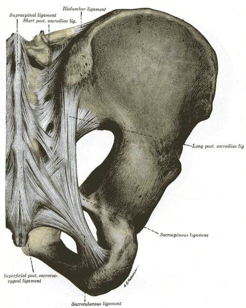
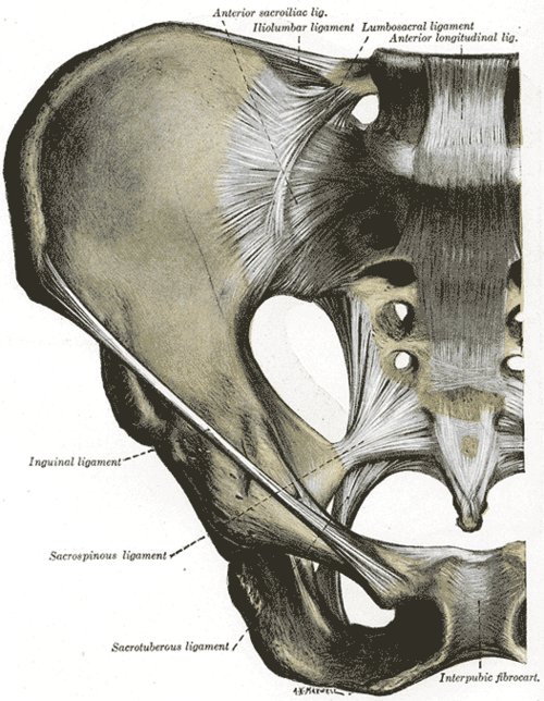
Two strong ligaments from the sacrum and coccyx to the ischium that stabilize the sacrum and turn the sciatic notches into the greater and lesser sciatic foramina, the gateways between pelvis, gluteal region and perineum.
Core anatomy
- Location
Posterolateral wall of the bony pelvis, deep to gluteus maximus.
- Attachments
Sacrotuberous: from the PSIS, the lateral border of the lower sacrum and coccyx and the coccyx to the medial margin of the ischial tuberosity. Sacrospinous: from the lateral margin of the lower sacrum and coccyx to the ischial spine; it lies anterior (deep) to the sacrotuberous ligament and is fused with the coccygeus muscle.
- Function
Resist upward rotation of the lower sacrum (and forward tilting of the upper sacrum) as body weight is transmitted through the sacroiliac joint. Together they convert the greater and lesser sciatic notches into the greater and lesser sciatic foramina.
- Relations
Greater sciatic foramen (above the sacrospinous ligament): piriformis passes through and divides it. Above piriformis: superior gluteal nerve and vessels. Below piriformis: inferior gluteal nerve and vessels, sciatic nerve, posterior femoral cutaneous nerve, nerve to quadratus femoris, and the pudendal nerve, internal pudendal vessels and nerve to obturator internus. Lesser sciatic foramen (between the two ligaments): tendon of obturator internus, and the pudendal nerve, internal pudendal vessels and nerve to obturator internus, which cross the ischial spine/sacrospinous ligament to re-enter the perineum.
- Notes
Mnemonic "PIN": the Pudendal nerve, Internal pudendal vessels and Nerve to obturator internus leave through the greater and re-enter through the lesser sciatic foramen.
Forms a boundary of
Mentioned in
Generated automatically from links in other entries.


Clinical correlations
Pudendal nerve block PANCE
- Mechanism / lesion
The pudendal nerve crosses the posterior surface of the sacrospinous ligament just medial to the ischial spine. Transvaginally, the palpable ischial spine guides the needle through the ligament.
- Classic presentation
Anesthesia of most of the perineum for the second stage of labor, operative vaginal delivery or perineal repair. Aspirate first: the internal pudendal vessels lie alongside.
Sacrospinous ligament fixation
- Mechanism / lesion
Surgical suspension of the vaginal apex to the sacrospinous ligament to treat apical (vault) prolapse.
- Classic presentation
Sutures placed too close to the ischial spine risk the pudendal nerve and internal pudendal vessels; placed too superiorly they risk the inferior gluteal vessels and sacral plexus, causing buttock pain.
Pudendal nerve entrapment
- Mechanism / lesion
Compression of the pudendal nerve between the sacrotuberous and sacrospinous ligaments or in the pudendal canal (e.g. long-distance cycling).
- Classic presentation
Burning perineal or genital pain that is worse sitting and relieved by standing or lying down.
High-yield PANCE pearls
- Sacrospinous → ischial spine; sacrotuberous → ischial tuberosity.
- PIN (pudendal nerve, internal pudendal vessels, nerve to obturator internus) exit the greater and enter the lesser sciatic foramen.
- Pudendal block: needle through the sacrospinous ligament just medial to the ischial spine.
Imaging anatomy
- CT
CT shows ossification or avulsion at their attachments and guides pudendal nerve injections near the ischial spine.
- MRI
MRI shows the ligaments as low-signal bands and the pudendal canal; used in suspected pudendal nerve entrapment.
Physical exam
- Surface landmarks
The ischial spine is palpable on vaginal (or rectal) exam at about 4 and 8 o'clock; the sacrotuberous ligament can be felt deep in the buttock between the ischial tuberosity and the sacrum.
- Palpation
Tenderness over the ischial spine or along the sacrospinous ligament on internal exam supports pudendal nerve entrapment.
Self-quiz
During a transvaginal pudendal nerve block, which palpable landmark guides needle placement?
Correct answer: B
The pudendal nerve crosses the sacrospinous ligament just medial to the ischial spine, which is palpable through the lateral vaginal wall. The needle passes through the ligament next to the spine.
Show answer
B
The pudendal nerve crosses the sacrospinous ligament just medial to the ischial spine, which is palpable through the lateral vaginal wall. The needle passes through the ligament next to the spine.
Which structure passes through the lesser sciatic foramen?
Correct answer: C
The obturator internus tendon leaves the pelvis through the lesser sciatic foramen, along with the pudendal nerve, internal pudendal vessels and nerve to obturator internus entering the perineum. The others pass through the greater sciatic foramen.
Show answer
C
The obturator internus tendon leaves the pelvis through the lesser sciatic foramen, along with the pudendal nerve, internal pudendal vessels and nerve to obturator internus entering the perineum. The others pass through the greater sciatic foramen.
The sacrospinous ligament attaches laterally to which structure?
Correct answer: B
The sacrospinous ligament runs to the ischial spine; the sacrotuberous ligament runs to the ischial tuberosity.
Show answer
B
The sacrospinous ligament runs to the ischial spine; the sacrotuberous ligament runs to the ischial tuberosity.
The sacrotuberous ligament runs from the sacrum and coccyx to which landmark?
Correct answer: E
Sacrotuberous → ischial tuberosity; sacrospinous → ischial spine. Together they convert the sciatic notches into the greater and lesser sciatic foramina and resist upward rotation of the lower sacrum.
Show answer
E
Sacrotuberous → ischial tuberosity; sacrospinous → ischial spine. Together they convert the sciatic notches into the greater and lesser sciatic foramina and resist upward rotation of the lower sacrum.
Which nerve leaves the pelvis through the greater sciatic foramen and then re-enters through the lesser sciatic foramen?
Correct answer: C
Mnemonic PIN: the Pudendal nerve, Internal pudendal vessels and Nerve to obturator internus exit through the greater sciatic foramen below piriformis and re-enter through the lesser sciatic foramen. The sciatic, inferior gluteal and posterior femoral cutaneous nerves stay in the gluteal region; the superior gluteal nerve exits above piriformis.
Show answer
C
Mnemonic PIN: the Pudendal nerve, Internal pudendal vessels and Nerve to obturator internus exit through the greater sciatic foramen below piriformis and re-enter through the lesser sciatic foramen. The sciatic, inferior gluteal and posterior femoral cutaneous nerves stay in the gluteal region; the superior gluteal nerve exits above piriformis.