Rhomboid major and minor
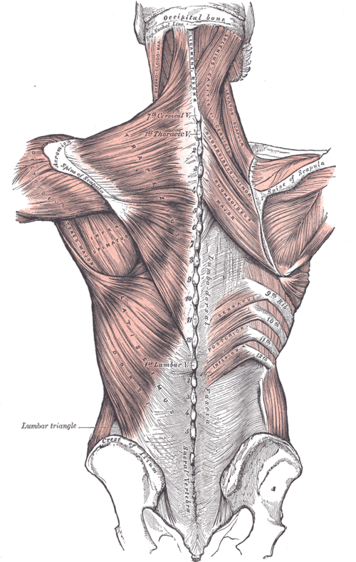
Two flat muscles between the upper thoracic spine and the medial border of the scapula that retract the scapula. They are supplied by the dorsal scapular nerve (C5).
Core anatomy
- Location
Deep to trapezius, between the vertebral column and the medial border of the scapula.
- Group
Superficial extrinsic back muscles (deep to trapezius), with levator scapulae.
- Origin
Minor: nuchal ligament and spinous processes of C7–T1. Major: spinous processes of T2–T5.
- Insertion
Medial border of the scapula: minor at the level of the scapular spine; major from the spine to the inferior angle.
- Innervation
Dorsal scapular nerve (C5, often with C4), from the C5 root of the brachial plexus.
- Blood supply
Dorsal scapular artery (or the deep branch of the transverse cervical artery) and posterior intercostal branches.
- Action
- Retract the scapula (squeeze the shoulder blades together)
- Rotate the glenoid downward; hold the scapula against the thoracic wall
- Relations
The dorsal scapular nerve pierces the middle scalene and descends deep to levator scapulae and the rhomboids with the dorsal scapular artery.

Clinical correlations
Dorsal scapular nerve injury or entrapment
- Mechanism / lesion
Entrapment in the middle scalene, or injury in the neck.
- Classic presentation
Aching along the medial border of the scapula, weak retraction, and mild winging of the medial border (less marked than with serratus paralysis).
Interscapular (rhomboid) muscle pain
- Mechanism / lesion
Postural strain and overuse. Interscapular pain can also be referred from cervical disease, gallbladder, or thoracic aortic pathology.
- Classic presentation
Aching between the shoulder blades with tender trigger points; always consider referred causes.
High-yield PANCE pearls
- Rhomboids: retraction; dorsal scapular nerve (C5) from the root.
- Scapular winging: medial (long thoracic/serratus) vs lateral (CN XI/trapezius); rhomboid weakness gives only mild medial-border prominence.
Imaging anatomy
- MRI
Muscle atrophy with chronic denervation.
- Key views & landmarks
- EMG for dorsal scapular neuropathy
Physical exam
- Surface landmarks
Between the medial borders of the scapulae and the spinous processes of C7–T5.
- Strength / function testing
Resisted retraction — hands on hips, the patient pushes the elbows backward against resistance while the examiner palpates the rhomboids medial to the scapula.
Self-quiz
Which nerve supplies the rhomboid muscles?
Correct answer: B
The dorsal scapular nerve (C5), arising directly from the C5 root, supplies both rhomboids and levator scapulae (with C3–C4).
Show answer
B
The dorsal scapular nerve (C5), arising directly from the C5 root, supplies both rhomboids and levator scapulae (with C3–C4).
What is the main action of the rhomboids?
Correct answer: B
The rhomboids retract the scapula and rotate the glenoid downward. Serratus anterior protracts and upwardly rotates.
Show answer
B
The rhomboids retract the scapula and rotate the glenoid downward. Serratus anterior protracts and upwardly rotates.
Rhomboid major attaches to the spinous processes of which vertebrae?
Correct answer: C
Rhomboid major arises from T2–T5; rhomboid minor from C7–T1 (and the nuchal ligament).
Show answer
C
Rhomboid major arises from T2–T5; rhomboid minor from C7–T1 (and the nuchal ligament).