Temporomandibular joint
Synovial joint between the mandibular condyle and the temporal bone, divided by an articular disc into two compartments that allow hinging and gliding. TMJ disorders and anterior dislocation are common.
Core anatomy
- Location
Just in front of the tragus of the ear.
- Classification
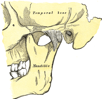
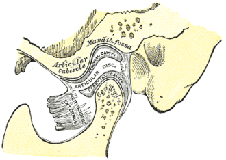
Synovial joint (modified hinge with gliding), divided by an articular disc into upper and lower compartments
- Articulating surfaces
Condyle of the mandible and the mandibular fossa and articular tubercle of the temporal bone (skull)
- Capsule
Loose; the articular surfaces are covered by fibrocartilage (not hyaline).
- Ligaments
- Lateral (temporomandibular) ligament: main stabilizer; prevents posterior displacement
- Sphenomandibular and stylomandibular ligaments (accessory)
- Articular disc: fibrocartilage, attached to the capsule and to the lateral pterygoid anteriorly
- Movements
- Lower compartment: hinge (rotation) — first part of mouth opening
- Upper compartment: gliding (translation) of condyle and disc forward onto the articular tubercle — wide opening, protrusion
- Side-to-side (grinding) movements
- Muscles: see muscles of mastication
- Stability
Most stable with the teeth together (occlusion). Wide opening moves the condyle onto the articular tubercle, the position from which it can dislocate.
- Innervation
Auriculotemporal and masseteric branches of the mandibular nerve (V3; trigeminal nerve (CN V)).
- Blood supply
Superficial temporal and maxillary arteries (branches of the external carotid artery).
- Relations
The parotid gland lies behind and lateral to the joint; the external acoustic meatus lies directly behind the condyle, so TMJ pain is often felt as ear pain.
Mentioned in
Generated automatically from links in other entries.


Clinical correlations
Temporomandibular disorders PANCE
- Mechanism / lesion
Myofascial pain of the masticatory muscles, disc displacement or degenerative joint disease; associated with bruxism, clenching and stress.
- Classic presentation
Jaw or preauricular pain (often felt as ear pain with a normal otoscopic exam), clicking, limited opening, headache; worse with chewing. Managed conservatively (soft diet, NSAIDs, night guard, physical therapy).
TMJ dislocation
- Mechanism / lesion
Anterior dislocation during wide yawning or opening; the condyle locks in front of the articular tubercle, held by muscle spasm.
- Classic presentation
Open-locked jaw, unable to close, prominent chin. Reduced by pressing down and back on the lower molars (protect the thumbs).
High-yield PANCE pearls
- Ear pain with a normal ear exam, jaw clicking, worse with chewing → TMJ disorder.
- Jaw stuck open after a yawn → anterior TMJ dislocation.
- TMJ has an articular disc and fibrocartilage surfaces.
Imaging anatomy
- X-ray
Panoramic radiograph for gross bony changes.
- CT
Bony anatomy, fractures and ankylosis.
- MRI
MRI (open and closed mouth) shows the articular disc position and effusions — the test for internal derangement.
Physical exam
- Surface landmarks
The condyle is palpable just in front of the tragus, moving forward as the mouth opens.
- Palpation
Palpate the joint (in front of the tragus or via the external ear canal) during opening and closing for tenderness, clicking or crepitus; palpate masseter and temporalis.
- Strength / function testing
Measure maximal interincisal opening (normal about 40–50 mm, roughly three fingers' breadth); observe deviation of the jaw on opening (toward the affected side). verify
Self-quiz
A 32-year-old woman has several months of left ear pain, worse when chewing, with clicking of the jaw. She grinds her teeth at night. Otoscopy is normal. What is the most likely diagnosis?
Correct answer: B
Temporomandibular disorder commonly causes ear/preauricular pain with a normal ear exam, clicking and pain on chewing, often with bruxism. Trigeminal neuralgia causes brief electric paroxysms.
Show answer
B
Temporomandibular disorder commonly causes ear/preauricular pain with a normal ear exam, clicking and pain on chewing, often with bruxism. Trigeminal neuralgia causes brief electric paroxysms.
Which structure divides the TMJ into upper and lower compartments?
Correct answer: B
The fibrocartilaginous articular disc divides the joint into an upper (gliding) and lower (hinge) compartment.
Show answer
B
The fibrocartilaginous articular disc divides the joint into an upper (gliding) and lower (hinge) compartment.
A man yawns widely and now can't close his mouth; his chin juts forward. What has happened, and how is it reduced?
Correct answer: C
Wide opening glides the condyle onto the articular tubercle; in an anterior dislocation it locks in front of it, held by muscle spasm. Reduce it by pressing down and back on the lower molars, protecting the thumbs.
Show answer
C
Wide opening glides the condyle onto the articular tubercle; in an anterior dislocation it locks in front of it, held by muscle spasm. Reduce it by pressing down and back on the lower molars, protecting the thumbs.