Phrenic nerve
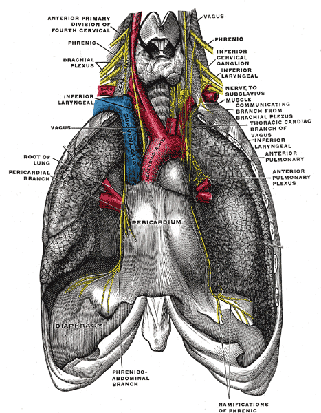
Nerve from C3–C5 that descends through the neck and mediastinum. It is the sole motor supply of the diaphragm and carries sensation from the pericardium, mediastinal pleura and central diaphragm.
Core anatomy
- Location
Neck (on anterior scalene) → superior mediastinum → middle mediastinum (on the pericardium) → diaphragm.
- Root values
C3, C4, C5 (mainly C4)
- Origin
Ventral rami of C3–C5 (cervical plexus, with a contribution from C5).
- Course
Descends on the anterior surface of anterior scalene, deep to the prevertebral fascia, crossing it from lateral to medial. Enters the thorax between the subclavian artery and vein. In the thorax both nerves pass anterior to the root of the lung (the vagus nerves pass posterior), running between the fibrous pericardium and the mediastinal pleura with the pericardiacophrenic vessels. The right phrenic nerve runs along the right brachiocephalic vein, superior vena cava and right atrium and pierces the diaphragm near the caval opening. The left phrenic nerve descends over the arch of the aorta and the left ventricle and pierces the muscular diaphragm to the left of the pericardium.
- Branches
- Motor branches to the diaphragm (entering from its abdominal surface)
- Sensory branches to the fibrous and parietal pericardium, mediastinal and diaphragmatic pleura, and central diaphragmatic peritoneum
- Motor
Diaphragm, its only motor supply.
- Sensory
Pericardium, mediastinal pleura, and the central parts of the diaphragmatic pleura and peritoneum. Pain from these sites is referred to the C3–C5 dermatomes (shoulder and neck).
- Relations
Accompanied in the thorax by the pericardiacophrenic artery and vein (from the internal thoracic artery). Anterior to the lung root; the vagus passes posterior to it.
Muscles innervated
- Diaphragm — Motor: phrenic nerve (C3, C4, C5), the sole motor supply. Sensory: phrenic nerve to the central part; the lower intercostal and subcostal nerves to the peripheral costal part.
Found in
Mentioned in
Generated automatically from links in other entries.

Clinical correlations
Phrenic nerve palsy PANCE
- Mechanism / lesion
Injury during cardiac or thoracic surgery (including cold cardioplegia), invasion by lung cancer or mediastinal tumors, neck trauma or surgery, interscalene brachial plexus block (ipsilateral phrenic blockade is very common), and herpes zoster or neuralgic amyotrophy.
- Classic presentation
Elevated hemidiaphragm on CXR. Unilateral paralysis may be asymptomatic or cause exertional dyspnea. Bilateral paralysis causes orthopnea and respiratory failure.
Referred shoulder pain PANCE
- Mechanism / lesion
Irritation of the diaphragm, pericardium or mediastinal pleura (e.g. pericarditis, hemoperitoneum, subphrenic abscess).
- Classic presentation
Pain at the shoulder or neck, which share the C3–C5 dermatomes. Example: Kehr sign (left shoulder pain) with splenic rupture.
High cervical spinal cord injury PANCE
- Mechanism / lesion
Injury at or above C3–C5 disrupts phrenic motor neurons.
- Classic presentation
Loss of diaphragmatic breathing. Injuries above about C3 usually need long-term ventilatory support; partial C4–C5 injuries may leave some function.
High-yield PANCE pearls
- "C3, 4, 5 keeps the diaphragm alive."
- Phrenic nerve passes anterior to the lung root; vagus passes posterior.
- Interscalene block → ipsilateral diaphragmatic paresis; use caution in patients with significant lung disease.
- New elevated hemidiaphragm in a smoker → consider lung cancer involving the phrenic nerve.
Imaging anatomy
- X-ray
The nerve isn't visible. An elevated hemidiaphragm is the indirect sign of palsy.
- CT
CT chest looks for a cause (mediastinal mass, lung cancer) along the course of the nerve.
- Ultrasound
Diaphragmatic ultrasound (M-mode excursion, thickening fraction) diagnoses paralysis at the bedside. The phrenic nerve itself can be seen on anterior scalene in the neck with high-frequency ultrasound.
- Key views & landmarks
- Fluoroscopic or ultrasound sniff test: paradoxical upward movement of the paralyzed side
Physical exam
- Surface landmarks
In the neck, lies on anterior scalene deep to sternocleidomastoid.
- Strength / function testing
Observe for paradoxical abdominal motion (inward on inspiration) when supine in bilateral paralysis. Percuss for reduced diaphragmatic excursion on the affected side.
Self-quiz
Shortly after an interscalene brachial plexus block for shoulder surgery, a patient with COPD becomes dyspneic. Chest radiograph shows a new elevation of the ipsilateral hemidiaphragm. Which nerve has been affected?
Correct answer: B
The phrenic nerve lies on anterior scalene, next to the interscalene groove, and is almost always blocked by an interscalene injection. In patients with limited respiratory reserve this can cause significant dyspnea.
Show answer
B
The phrenic nerve lies on anterior scalene, next to the interscalene groove, and is almost always blocked by an interscalene injection. In patients with limited respiratory reserve this can cause significant dyspnea.
Which relationship of the phrenic nerve to the root of the lung is correct?
Correct answer: B
In the mediastinum the phrenic nerve passes anterior to the lung root, between the pericardium and pleura. The vagus passes posterior to it, contributing to the pulmonary and esophageal plexuses.
Show answer
B
In the mediastinum the phrenic nerve passes anterior to the lung root, between the pericardium and pleura. The vagus passes posterior to it, contributing to the pulmonary and esophageal plexuses.
A patient with acute pericarditis reports sharp chest pain that also radiates to the trapezius ridge. Which nerve carries this pain?
Correct answer: B
The pericardium is innervated by the phrenic nerve (C3–C5). Pain from pericarditis radiating to the trapezius ridge is characteristic and reflects the shared C3–C5 dermatomes.
Show answer
B
The pericardium is innervated by the phrenic nerve (C3–C5). Pain from pericarditis radiating to the trapezius ridge is characteristic and reflects the shared C3–C5 dermatomes.
The phrenic nerve enters the thorax between which two structures?
Correct answer: D
The phrenic nerve descends on the anterior surface of anterior scalene and enters the thorax between the subclavian artery and vein. The roots of the brachial plexus pass between the anterior and middle scalenes.
Show answer
D
The phrenic nerve descends on the anterior surface of anterior scalene and enters the thorax between the subclavian artery and vein. The roots of the brachial plexus pass between the anterior and middle scalenes.
Which symptom is typical of bilateral diaphragmatic paralysis?
Correct answer: C
Bilateral paralysis causes orthopnea (the abdominal contents push the flaccid diaphragm up when supine), paradoxical inward movement of the abdomen on inspiration, and potentially respiratory failure. Unilateral paralysis is often asymptomatic.
Show answer
C
Bilateral paralysis causes orthopnea (the abdominal contents push the flaccid diaphragm up when supine), paradoxical inward movement of the abdomen on inspiration, and potentially respiratory failure. Unilateral paralysis is often asymptomatic.