Uterine tubes
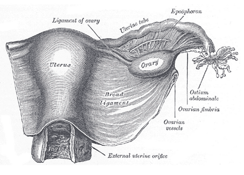
Paired tubes that carry the oocyte from the ovary to the uterus; fertilization usually occurs in the ampulla. They are the most common site of ectopic pregnancy and are damaged by pelvic inflammatory disease.
Core anatomy
- Location
In the free upper edge of the broad ligament (the mesosalpinx), from the uterine horns toward the lateral pelvic wall, arching over the ovary. About 10 cm long.
- Parts
- Infundibulum: funnel-shaped lateral end opening into the peritoneal cavity through the abdominal ostium, fringed by fimbriae (one, the ovarian fimbria, attaches to the ovary)
- Ampulla: the widest and longest part; the usual site of fertilization and of ectopic pregnancy
- Isthmus: narrow, thick-walled part near the uterus
- Uterine (intramural) part: passes through the myometrium to open at the uterine ostium; interstitial (cornual) ectopic pregnancies rupture late and bleed heavily
- Wall: ciliated and secretory (peg) columnar epithelium and smooth muscle, which move the oocyte toward the uterus
- Relations
The abdominal ostium makes the female peritoneal cavity communicate with the exterior (via tube, uterus and vagina) — a route for ascending infection. The ovary lies below and behind the tube.
- Arterial supply
Tubal branches of the uterine artery (medial) and the ovarian artery (lateral), which anastomose.
- Venous drainage
Veins parallel the arteries to the uterine and ovarian (pampiniform) venous plexuses.
- Lymphatic drainage
Mainly lumbar (para-aortic) nodes with the ovarian vessels.
- Innervation
Ovarian and uterine (uterovaginal) plexuses; visceral pain with sympathetic afferents to about T10–L1 verify
Mentioned in
Generated automatically from links in other entries.

Clinical correlations
Ectopic pregnancy PANCE
- Mechanism / lesion
Implantation outside the uterine cavity; about 95% are tubal, most in the ampulla. Risk factors: prior PID, prior ectopic, tubal surgery or ligation, pregnancy with an IUD in place, assisted reproduction, smoking.
- Classic presentation
Amenorrhea, abdominal/pelvic pain and vaginal bleeding, typically at 6–8 weeks; adnexal tenderness or mass. Rupture: severe pain, peritoneal signs, shoulder-tip pain, hemorrhagic shock.
- Notes
Positive β-hCG with no intrauterine pregnancy on TVUS is ectopic until proven otherwise (especially above the discriminatory level; ACOG advises a conservatively high cutoff, up to about 3,500 mIU/mL). Stable, unruptured, meeting criteria → methotrexate; unstable or ruptured → surgery (salpingostomy or salpingectomy). Give anti-D to Rh-negative women.
Pelvic inflammatory disease (salpingitis) PANCE
- Mechanism / lesion
Ascending polymicrobial infection, classically Chlamydia trachomatis and Neisseria gonorrhoeae.
- Classic presentation
Lower abdominal pain with cervical motion, uterine or adnexal tenderness (CDC minimum criteria), fever, discharge. Complications: tubo-ovarian abscess, infertility, ectopic pregnancy, chronic pelvic pain, Fitz-Hugh–Curtis perihepatitis (right upper quadrant pain).
- Notes
Outpatient treatment (CDC 2021): ceftriaxone IM + doxycycline + metronidazole for 14 days.
Tubal sterilization
- Mechanism / lesion
Ligation, occlusion or removal (salpingectomy) of the tubes; salpingectomy also lowers ovarian cancer risk, since many high-grade serous cancers begin in the fimbriae.
- Classic presentation
Permanent contraception; if pregnancy occurs afterward, it is more likely to be ectopic.
High-yield PANCE pearls
- Pregnant, pain + bleeding, no intrauterine pregnancy on TVUS → ectopic (ampulla most common).
- Stable unruptured ectopic → methotrexate; unstable → surgery.
- PID: cervical motion tenderness; chlamydia/gonorrhea; complications infertility and ectopic.
- PID + RUQ pain → Fitz-Hugh–Curtis syndrome.
- Fertilization occurs in the ampulla.
Imaging anatomy
- X-ray
Hysterosalpingography tests tubal patency in infertility (contrast spilling freely into the peritoneal cavity).
- Ultrasound
Transvaginal ultrasound with quantitative β-hCG: absence of an intrauterine gestational sac, an adnexal mass separate from the ovary ("ring of fire" on Doppler), and free fluid in the rectouterine pouch. Bedside FAST in an unstable patient. Tubo-ovarian abscess appears as a complex adnexal mass.
- Key views & landmarks
- Transvaginal ultrasound (adnexa and rectouterine pouch)
- Hysterosalpingogram for tubal patency
Physical exam
- Surface landmarks
The tubes are not normally palpable; adnexal fullness or tenderness is sought lateral to the uterus.
- Palpation
Bimanual exam — adnexal tenderness or mass, cervical motion tenderness ("chandelier sign").
- Strength / function testing
Pregnancy test in every woman of reproductive age with abdominal pain or abnormal bleeding.
Self-quiz
A 27-year-old woman with a history of PID has 6 weeks of amenorrhea, left lower quadrant pain and light vaginal bleeding. BP is 118/76 mm Hg. β-hCG is 2,800 mIU/mL, and transvaginal ultrasound shows an empty uterus and a small left adnexal mass. What is the most likely diagnosis?
Correct answer: B
A positive β-hCG with an empty uterus and an adnexal mass is an ectopic pregnancy; prior PID is the classic risk factor (tubal scarring). She is stable, so methotrexate may be an option if criteria are met.
Show answer
B
A positive β-hCG with an empty uterus and an adnexal mass is an ectopic pregnancy; prior PID is the classic risk factor (tubal scarring). She is stable, so methotrexate may be an option if criteria are met.
What is the most common site of an ectopic pregnancy?
Correct answer: C
About 95% of ectopic pregnancies are tubal, and most of these are in the ampulla, which is also where fertilization normally occurs.
Show answer
C
About 95% of ectopic pregnancies are tubal, and most of these are in the ampulla, which is also where fertilization normally occurs.
A 22-year-old woman with PID treated last month now has right upper quadrant pain that is worse with breathing. Liver tests are normal. What is the most likely cause?
Correct answer: B
Fitz-Hugh–Curtis syndrome is perihepatitis (inflammation of the liver capsule, "violin-string" adhesions) after PID; the infection spreads from the open abdominal ostium of the tube through the peritoneal cavity. Liver tests are usually normal.
Show answer
B
Fitz-Hugh–Curtis syndrome is perihepatitis (inflammation of the liver capsule, "violin-string" adhesions) after PID; the infection spreads from the open abdominal ostium of the tube through the peritoneal cavity. Liver tests are usually normal.
A hemodynamically stable woman has an unruptured tubal ectopic pregnancy and meets the criteria for medical management. What is the most appropriate treatment?
Correct answer: E
A stable, unruptured ectopic pregnancy that meets criteria is treated with methotrexate; an unstable or ruptured ectopic needs surgery (salpingostomy or salpingectomy). Rh-negative women should receive anti-D.
Show answer
E
A stable, unruptured ectopic pregnancy that meets criteria is treated with methotrexate; an unstable or ruptured ectopic needs surgery (salpingostomy or salpingectomy). Rh-negative women should receive anti-D.
Why does salpingectomy for sterilization also lower the risk of ovarian cancer?
Correct answer: A
Many high-grade serous "ovarian" cancers originate in the fimbriae of the uterine tube, so removing the tubes (salpingectomy) lowers ovarian cancer risk as well as providing permanent contraception.
Show answer
A
Many high-grade serous "ovarian" cancers originate in the fimbriae of the uterine tube, so removing the tubes (salpingectomy) lowers ovarian cancer risk as well as providing permanent contraception.
Flashcards
See also
- Ovary
- Uterus
- Broad ligament
- Rectouterine pouch
- Lymph drainage of the pelvis and perineum (comparison table)
- Collapse in early pregnancy (clinical case)