Popliteal artery
Continuation of the femoral artery through the popliteal fossa, the deepest structure there. It is tethered and easily injured in knee dislocations and is the most common site of peripheral aneurysms.
Core anatomy
- Location
Popliteal fossa, lying deepest (against the femur and knee capsule).
- Origin
Continuation of the femoral artery at the adductor hiatus.
- Course
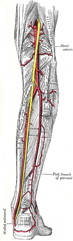
Descends through the popliteal fossa, deep to the popliteal vein and tibial nerve, and ends at the lower border of popliteus by dividing into the anterior and posterior tibial arteries.
- Branches
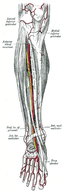
- Genicular arteries (superior medial and lateral, middle, inferior medial and lateral) forming the genicular anastomosis
- Sural arteries to gastrocnemius and soleus
- Anterior tibial artery: through the interosseous membrane into the anterior compartment; continues on the dorsum of the foot as the dorsalis pedis artery
- Posterior tibial artery (tibioperoneal trunk): deep posterior compartment, gives the fibular (peroneal) artery; passes behind the medial malleolus and divides into the medial and lateral plantar arteries
- Supplies
The knee joint and all of the leg and foot.
- Anastomoses
The genicular anastomosis around the knee connects with the descending genicular (femoral) and circumflex femoral arteries — insufficient to keep the leg alive after acute occlusion.
- Termination
Divides into the anterior tibial and posterior tibial arteries at the lower border of popliteus.
Structures supplied
Found in
Mentioned in
Generated automatically from links in other entries.


Clinical correlations
Popliteal artery injury in knee dislocation PANCE
- Mechanism / lesion
The artery is tethered above (adductor hiatus) and below (soleus arch), so knee dislocation (tibiofemoral) stretches or tears it — often an intimal tear with thrombosis.
- Classic presentation
Pulses may be present initially. Check pulses and the ankle–brachial index in every knee dislocation (ABI below 0.9 → CT angiography); ischemia beyond about 6–8 hours often leads to amputation. The common fibular nerve is often injured too.
Popliteal artery aneurysm PANCE
- Mechanism / lesion
The most common peripheral aneurysm, usually atherosclerotic in older men; frequently bilateral and associated with abdominal aortic aneurysm.
- Classic presentation
A pulsatile mass behind the knee; complications are thrombosis or distal embolism causing acute limb ischemia (rupture is rare). Image the other knee and the aorta.
Popliteal artery entrapment syndrome
- Mechanism / lesion
Abnormal relationship with the medial head of gastrocnemius compressing the artery; young athletes.
- Classic presentation
Exertional calf claudication in a young person; pulses diminish with active plantarflexion.
High-yield PANCE pearls
- Knee dislocation → check pulses and ABI → CTA if abnormal (popliteal artery injury).
- Pulsatile popliteal mass → popliteal aneurysm → check the other side and the aorta.
- Dorsalis pedis = continuation of the anterior tibial; posterior tibial pulse behind the medial malleolus.
- Deepest structure in the popliteal fossa = popliteal artery.
Imaging anatomy
- CT
CT angiography after knee dislocation with abnormal ABI or pulses, and for aneurysm planning.
- Ultrasound
Duplex ultrasound for aneurysm size and thrombus, and entrapment (with provocative maneuvers).
- Key views & landmarks
- Ankle–brachial index (screening after knee dislocation)
- CT angiography of the lower extremity
Physical exam
- Surface landmarks
The popliteal pulse is felt deep in the popliteal fossa with the knee flexed about 30° and the muscles relaxed (press against the back of the tibia/femur).
- Palpation
An easily palpable, broad popliteal pulse suggests an aneurysm. Dorsalis pedis lateral to the EHL tendon on the dorsum; posterior tibial behind the medial malleolus.
- Strength / function testing
ABI; distal capillary refill; motor/sensory exam of the foot.
Self-quiz
A 25-year-old man has a knee dislocation after a motorcycle crash; it is reduced in the ED. His dorsalis pedis pulse is palpable but weaker than on the left. What is the most appropriate next step?
Correct answer: B
The popliteal artery is tethered and often injured in knee dislocations; a palpable pulse doesn't exclude an intimal tear. An ABI below 0.9 mandates CT angiography (or surgical exploration if the limb is ischemic).
Show answer
B
The popliteal artery is tethered and often injured in knee dislocations; a palpable pulse doesn't exclude an intimal tear. An ABI below 0.9 mandates CT angiography (or surgical exploration if the limb is ischemic).
A 72-year-old man has a pulsatile mass behind his right knee. What other study is most important?
Correct answer: B
Popliteal aneurysms are often bilateral and frequently associated with abdominal aortic aneurysm, so both should be screened.
Show answer
B
Popliteal aneurysms are often bilateral and frequently associated with abdominal aortic aneurysm, so both should be screened.
Why is the popliteal artery so often injured in knee dislocations?
Correct answer: C
The popliteal artery is fixed above (adductor hiatus) and below (soleal arch), so tibiofemoral dislocation stretches or tears it, often as an intimal tear with thrombosis. It is also the deepest structure in the fossa, against the femur and capsule.
Show answer
C
The popliteal artery is fixed above (adductor hiatus) and below (soleal arch), so tibiofemoral dislocation stretches or tears it, often as an intimal tear with thrombosis. It is also the deepest structure in the fossa, against the femur and capsule.
The dorsalis pedis artery is the continuation of which artery?
Correct answer: A
The anterior tibial artery passes through the interosseous membrane into the anterior compartment and continues on the dorsum of the foot as the dorsalis pedis. The posterior tibial pulse is felt behind the medial malleolus.
Show answer
A
The anterior tibial artery passes through the interosseous membrane into the anterior compartment and continues on the dorsum of the foot as the dorsalis pedis. The posterior tibial pulse is felt behind the medial malleolus.
A 22-year-old runner has calf claudication with exercise. His pedal pulses weaken when he actively plantarflexes against resistance. What is the most likely diagnosis?
Correct answer: E
In popliteal artery entrapment, an abnormal relationship with the medial head of gastrocnemius compresses the artery, causing exertional claudication in young athletes, with pulses that diminish during active plantarflexion.
Show answer
E
In popliteal artery entrapment, an abnormal relationship with the medial head of gastrocnemius compresses the artery, causing exertional claudication in young athletes, with pulses that diminish during active plantarflexion.