Medulla oblongata
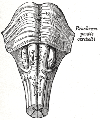
Lowest part of the brainstem, continuous with the spinal cord at the foramen magnum. It contains the cardiorespiratory centers, the nuclei of CN IX–XII and the pyramidal decussation; PICA strokes cause the lateral medullary (Wallenberg) syndrome.
Core anatomy
- Location
From the lower border of the pons to the foramen magnum, where it becomes the spinal cord; in front of the cerebellum and the lower fourth ventricle.
- Parts
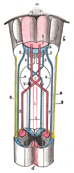
- Pyramids (ventral): corticospinal tracts; about 85–90% cross in the pyramidal decussation at the medulla–cord junction (see corticospinal tract)
- Olives (inferior olivary nucleus → cerebellum via the inferior cerebellar peduncle)
- Nuclei: hypoglossal (XII), dorsal motor nucleus of X, nucleus ambiguus (IX, X — swallowing, voice), nucleus of the solitary tract (taste, visceral afferents), spinal trigeminal nucleus and tract (ipsilateral face pain/temperature), vestibular nuclei, gracile and cuneate nuclei (dorsal columns → sensory decussation → medial lemniscus)
- Cardiovascular and respiratory centers (reticular formation); area postrema — chemoreceptor trigger zone for vomiting (outside the blood–brain barrier)
- Descending sympathetic fibers in the lateral medulla (lesion → Horner syndrome)
- Relations
The cerebellar tonsils lie just above the foramen magnum behind the medulla.
- Arterial supply
Vertebral arteries, PICA (lateral medulla), anterior spinal artery (medial medulla) — see vertebrobasilar system.
- Venous drainage
Medullary veins → inferior petrosal sinus and spinal veins.
- Notes
CN IX, X (postolivary sulcus), XI (lateral), XII (preolivary sulcus) emerge here — see glossopharyngeal nerve (CN IX), vagus nerve (CN X), accessory nerve (CN XI), hypoglossal nerve (CN XII).
Mentioned in
- Cerebellum
- Corticospinal tract
- Dorsal column–medial lemniscus pathway
- Glossopharyngeal nerve (CN IX)
- Hypoglossal nerve (CN XII)
- Pons
- Spinothalamic tract
- Vagus nerve (CN X)
- Vertebrobasilar system
Generated automatically from links in other entries.
Swipe sideways to see the whole diagram →


Clinical correlations
Lateral medullary (Wallenberg) syndrome PANCE
- Mechanism / lesion
Occlusion of the vertebral artery or PICA (dissection, atherosclerosis).
- Classic presentation
Ipsilateral loss of pain/temperature on the face (spinal trigeminal) with contralateral loss on the body (spinothalamic); dysphagia, hoarseness, reduced gag (nucleus ambiguus); ipsilateral Horner syndrome; vertigo, nystagmus, nausea (vestibular); ipsilateral limb ataxia; hiccups. No limb weakness.
Medial medullary syndrome PANCE
- Mechanism / lesion
Occlusion of the anterior spinal artery or vertebral paramedian branches.
- Classic presentation
Contralateral hemiparesis (pyramid), contralateral loss of proprioception/vibration (medial lemniscus), ipsilateral tongue deviation (XII).
Tonsillar herniation and Chiari I malformation PANCE
- Mechanism / lesion
Cerebellar tonsils pushed through the foramen magnum, compressing the medulla (raised ICP, posterior fossa mass, or lumbar puncture with a mass lesion); Chiari I — congenital tonsillar descent.
- Classic presentation
Herniation: neck stiffness, Cushing triad (hypertension, bradycardia, irregular respirations), then respiratory arrest. Chiari I: occipital headache worse with cough/Valsalva, syringomyelia (cape-like loss of pain/temperature).
High-yield PANCE pearls
- Ipsilateral face + contralateral body pain/temp loss + dysphagia/hoarseness + Horner → Wallenberg (PICA/vertebral).
- Contralateral hemiparesis + contralateral proprioception loss + ipsilateral tongue → medial medullary (anterior spinal).
- Cushing triad: hypertension, bradycardia, irregular breathing → impending herniation.
- Medulla = CN IX, X, XI, XII + cardiorespiratory centers.
- Area postrema = chemoreceptor trigger zone (vomiting).
Imaging anatomy
- CT
CT for hemorrhage and signs of raised ICP; image before lumbar puncture when a mass is possible.
- MRI
MRI with DWI — medullary infarcts (often missed on CT); sagittal MRI for Chiari malformation and syrinx; MRA/CTA for vertebral dissection.
Physical exam
- Strength / function testing
Palate elevation, gag, voice and swallow (IX, X), tongue (XII), SCM/trapezius (XI); facial and body pinprick/temperature (crossed sensory loss), pupils and ptosis (Horner), limb coordination, nystagmus.
Self-quiz
A 48-year-old has sudden vertigo, hoarseness and difficulty swallowing. Exam shows a small left pupil with ptosis, reduced pain sensation on the left face and the right side of the body, and left limb ataxia. Strength is normal. Which artery is occluded?
Correct answer: B
Lateral medullary (Wallenberg) syndrome from left PICA/vertebral occlusion: crossed pain/temperature loss, nucleus ambiguus signs (dysphagia, hoarseness), ipsilateral Horner and ataxia, with no weakness.
Show answer
B
Lateral medullary (Wallenberg) syndrome from left PICA/vertebral occlusion: crossed pain/temperature loss, nucleus ambiguus signs (dysphagia, hoarseness), ipsilateral Horner and ataxia, with no weakness.
A patient with a large cerebellar hemorrhage becomes hypertensive with a heart rate of 44/min and irregular breathing. What is happening?
Correct answer: B
The Cushing triad signals raised ICP with brainstem (medullary) compression — impending tonsillar herniation. Posterior fossa masses need urgent neurosurgical decompression.
Show answer
B
The Cushing triad signals raised ICP with brainstem (medullary) compression — impending tonsillar herniation. Posterior fossa masses need urgent neurosurgical decompression.
The area postrema of the medulla has which function?
Correct answer: A
The area postrema lies outside the blood–brain barrier and acts as the chemoreceptor trigger zone for vomiting.
Show answer
A
The area postrema lies outside the blood–brain barrier and acts as the chemoreceptor trigger zone for vomiting.
A 30-year-old has occipital headaches worse with coughing and a cape-like loss of pain and temperature over the shoulders. MRI shows the cerebellar tonsils below the foramen magnum. What is the diagnosis?
Correct answer: E
Chiari I malformation is congenital descent of the cerebellar tonsils, causing occipital headache with cough/Valsalva and often a syrinx (cape-like pain/temperature loss).
Show answer
E
Chiari I malformation is congenital descent of the cerebellar tonsils, causing occipital headache with cough/Valsalva and often a syrinx (cape-like pain/temperature loss).
In lateral medullary syndrome, damage to which nucleus causes dysphagia and hoarseness?
Correct answer: C
The nucleus ambiguus supplies the pharyngeal and laryngeal muscles (CN IX, X), so its damage causes dysphagia, hoarseness and a reduced gag. The spinal trigeminal nucleus causes the ipsilateral face pain/temperature loss.
Show answer
C
The nucleus ambiguus supplies the pharyngeal and laryngeal muscles (CN IX, X), so its damage causes dysphagia, hoarseness and a reduced gag. The spinal trigeminal nucleus causes the ipsilateral face pain/temperature loss.