Tibial nerve
Larger terminal branch of the sciatic nerve, running through the popliteal fossa and deep posterior leg to the sole. It supplies the plantarflexors and inverters and the skin of the sole, and is compressed in the tarsal tunnel.
Core anatomy
- Location
Center of the popliteal fossa, the deep posterior compartment of the leg, then behind the medial malleolus into the sole.
- Root values
L4–S3.
- Origin
Sciatic nerve, at the apex of the popliteal fossa
- Course
Descends vertically through the popliteal fossa (the most superficial of the three main structures there: nerve, vein, artery), passes deep to the soleus arch, runs in the deep posterior compartment with the posterior tibial vessels, and passes behind the medial malleolus under the flexor retinaculum (tarsal tunnel), dividing into the medial and lateral plantar nerves.
- Branches
- Muscular: gastrocnemius, soleus, plantaris, popliteus (triceps surae and calcaneal tendon); tibialis posterior, flexor digitorum longus, flexor hallucis longus
- Medial sural cutaneous nerve → joins the fibular communicating branch to form the sural nerve (lateral foot and heel)
- Medial calcaneal branches (heel)
- Medial and lateral plantar nerves: intrinsic foot muscles and the skin of the sole
- Motor
Plantarflexion, inversion (tibialis posterior), toe flexion and the intrinsic muscles of the sole; also knee flexion (gastrocnemius, popliteus).
- Sensory
Sole of the foot (plantar nerves), heel (calcaneal branches), lateral foot (sural nerve, with a fibular contribution).
- Relations
Tarsal tunnel contents, anterior to posterior: tibialis posterior, flexor digitorum longus, posterior tibial artery and veins, tibial nerve, flexor hallucis longus ("Tom, Dick And Very Nervous Harry").
Muscles innervated
- Triceps surae and calcaneal tendon — tibial nerve (S1–S2)
Other structures innervated
Found in
Mentioned in
Generated automatically from links in other entries.
Swipe sideways to see the whole diagram →
Swipe sideways to see the whole diagram →
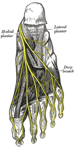

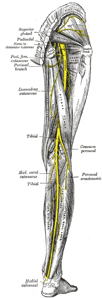

Clinical correlations
Tarsal tunnel syndrome PANCE
- Mechanism / lesion
Compression under the flexor retinaculum behind the medial malleolus (ganglia, varicosities, tenosynovitis, flat foot, trauma).
- Classic presentation
Burning pain, tingling and numbness of the sole and toes, worse with standing, walking or at night; Tinel sign behind the medial malleolus; the heel may be spared (calcaneal branches arise proximally).
Tibial nerve injury
- Mechanism / lesion
Posterior knee dislocation, popliteal trauma, deep posterior compartment syndrome.
- Classic presentation
Loss of plantarflexion and inversion, inability to stand on tiptoe, loss of the ankle reflex, numbness of the sole (risk of ulcers), a calcaneovalgus foot.
Sural nerve biopsy and grafts
- Mechanism / lesion
The sural nerve is purely sensory and easily found beside the small saphenous vein behind the lateral malleolus.
- Classic presentation
Used for nerve biopsy (e.g. vasculitic neuropathy) and as a nerve graft; leaves a numb patch on the lateral foot.
High-yield PANCE pearls
- Tibial nerve: plantarflexion + inversion, ankle reflex (S1), sole sensation.
- Burning sole pain with Tinel sign behind the medial malleolus → tarsal tunnel syndrome.
- Can't stand on tiptoe → tibial nerve (or S1/Achilles) problem.
Imaging anatomy
- MRI
Masses, ganglia or varicosities in the tarsal tunnel; MR neurography.
- Ultrasound
Shows compressive lesions in the tarsal tunnel and the nerve in the popliteal fossa.
Physical exam
- Surface landmarks
The tibial nerve and posterior tibial pulse lie midway between the medial malleolus and the Achilles tendon.
- Palpation
Tinel sign over the tarsal tunnel.
- Strength / function testing
Plantarflexion (tiptoe standing), inversion, toe flexion; ankle reflex (S1); sensation of the sole and heel. Nerve conduction studies confirm tarsal tunnel syndrome.
Self-quiz
A 50-year-old runner has burning pain and numbness on the sole of his left foot, worse at night. Tapping behind the medial malleolus reproduces the symptoms. Which nerve is compressed?
Correct answer: C
Tarsal tunnel syndrome is compression of the tibial nerve (or its plantar branches) under the flexor retinaculum behind the medial malleolus, causing burning and numbness of the sole with a positive Tinel sign.
Show answer
C
Tarsal tunnel syndrome is compression of the tibial nerve (or its plantar branches) under the flexor retinaculum behind the medial malleolus, causing burning and numbness of the sole with a positive Tinel sign.
A lesion of the tibial nerve in the popliteal fossa would cause loss of which movement?
Correct answer: C
The tibial nerve supplies the posterior leg muscles — plantarflexion (triceps surae) and inversion (tibialis posterior) — and the sole. Dorsiflexion and eversion are fibular nerve functions.
Show answer
C
The tibial nerve supplies the posterior leg muscles — plantarflexion (triceps surae) and inversion (tibialis posterior) — and the sole. Dorsiflexion and eversion are fibular nerve functions.
Which structure is the most posterior in the tarsal tunnel behind the medial malleolus?
Correct answer: E
From anterior to posterior: Tibialis posterior, flexor Digitorum longus, posterior tibial Artery and Veins, tibial Nerve, flexor Hallucis longus ("Tom, Dick And Very Nervous Harry").
Show answer
E
From anterior to posterior: Tibialis posterior, flexor Digitorum longus, posterior tibial Artery and Veins, tibial Nerve, flexor Hallucis longus ("Tom, Dick And Very Nervous Harry").
A purely sensory nerve, found beside the small saphenous vein behind the lateral malleolus, is commonly used for nerve biopsy. Which nerve is it?
Correct answer: C
The sural nerve (formed by the medial sural cutaneous branch of the tibial nerve and the fibular communicating branch) is purely sensory to the lateral foot and heel, so it is used for biopsy (e.g. vasculitic neuropathy) and as a nerve graft, leaving only a numb patch.
Show answer
C
The sural nerve (formed by the medial sural cutaneous branch of the tibial nerve and the fibular communicating branch) is purely sensory to the lateral foot and heel, so it is used for biopsy (e.g. vasculitic neuropathy) and as a nerve graft, leaving only a numb patch.
In tarsal tunnel syndrome, sensation over the heel is often preserved. Why?
Correct answer: B
The medial calcaneal branches of the tibial nerve arise before it passes under the flexor retinaculum, so heel sensation can be spared while the sole and toes (medial and lateral plantar nerves) are numb.
Show answer
B
The medial calcaneal branches of the tibial nerve arise before it passes under the flexor retinaculum, so heel sensation can be spared while the sole and toes (medial and lateral plantar nerves) are numb.