Cervical vertebrae
Seven small vertebrae of the neck with transverse foramina for the vertebral arteries. Cervical radiculopathy, myelopathy and trauma clearance are the key clinical topics.
Core anatomy
- Location
Neck, between the skull and T1. C1 and C2 are atypical (see atlas and axis (C1 and C2)).
- Classification
Irregular bones.
- Key features & landmarks
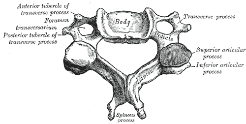
- Small, broad bodies with raised lateral lips (uncinate processes) forming uncovertebral joints (of Luschka) with the vertebra above
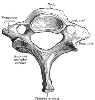
- Transverse foramina in every transverse process; the vertebral artery usually passes through those of C6–C1
- Bifid spinous processes (typically C3–C6)
- C7 (vertebra prominens): long, non-bifid spinous process, the first clearly palpable at the base of the neck
- Large triangular vertebral foramen (cervical enlargement of the cord)
- Anterior tubercle of C6 (carotid tubercle): the common carotid can be compressed against it
- Relations
Spinal nerves C1–C7 exit above the vertebra of the same number (C8 exits between C7 and T1), so a C5–C6 disc herniation affects the C6 root and C6–C7 affects C7. Facet joints are inclined at about 45°, allowing flexion, extension, lateral flexion and rotation.


Clinical correlations
Cervical radiculopathy PANCE
- Mechanism / lesion
Nerve root compression by disc herniation or foraminal narrowing (uncovertebral and facet osteophytes). C7 and C6 roots are most often involved.
- Classic presentation
Neck and arm pain in a dermatomal pattern with numbness, weakness and reduced reflexes. C5: deltoid, biceps reflex, lateral arm. C6: wrist extension, biceps/brachioradialis reflexes, thumb. C7: triceps, triceps reflex, middle finger. C8: finger flexion, little finger. Pain often eases with the hand on the head (shoulder abduction relief sign). Positive Spurling test.
Cervical spondylotic myelopathy PANCE
- Mechanism / lesion
Degenerative narrowing of the canal (disc bulge, osteophytes, ligamentum flavum hypertrophy, OPLL) compressing the spinal cord.
- Classic presentation
Clumsy hands, gait imbalance, upper motor neuron signs (hyperreflexia, Hoffmann sign, Babinski, clonus), Lhermitte sign. Progressive; MRI and surgical referral.
Cervical spine trauma clearance PANCE
- Mechanism / lesion
Decision rules identify alert trauma patients who don't need imaging.
- Classic presentation
NEXUS: imaging is not needed if there is no midline tenderness, no focal neurologic deficit, normal alertness, no intoxication, and no painful distracting injury. Canadian C-spine rule: high-risk factors (age 65 or more, dangerous mechanism, extremity paresthesias) mandate imaging; otherwise assess low-risk factors and active neck rotation. CT is the imaging test of choice in adults.
High-yield PANCE pearls
- C-roots exit above their vertebra: C5–C6 disc → C6 root; C6–C7 → C7 (most common radiculopathy).
- Root quick map: C5 deltoid/biceps reflex, C6 wrist extension/brachioradialis reflex/thumb, C7 triceps reflex/middle finger, C8 finger flexion/little finger.
- Spurling test → radiculopathy. Hoffmann sign, hyperreflexia → myelopathy.
- Trauma: NEXUS or Canadian C-spine rule; CT is first-line imaging in adults.
Imaging anatomy
- X-ray
Lateral view must show C1 through the C7–T1 junction (swimmer's view if needed); check the anterior vertebral, posterior vertebral and spinolaminar lines and prevertebral soft tissue. Obliques show foraminal narrowing. Largely replaced by CT in trauma.
- CT
First-line for cervical trauma in adults (fractures, alignment). CT angiography if vertebral artery injury is suspected (e.g. transverse foramen fracture).
- MRI
Discs, nerve roots, cord compression and signal change (myelopathy), ligamentous injury.
- Key views & landmarks
- CT cervical spine (trauma)
- MRI cervical spine (radiculopathy with red flags, myelopathy)
Physical exam
- Surface landmarks
C7 spinous process (vertebra prominens); the C6 carotid tubercle at about the level of the cricoid cartilage.
- Palpation
Midline tenderness (trauma), paraspinal spasm.
- Strength / function testing
Neck range of motion; myotomes, dermatomes and reflexes of the upper limbs; UMN signs (Hoffmann, Babinski, clonus).
Spurling test
- Technique
Extend the neck and rotate/laterally flex it toward the symptomatic side, then apply gentle axial compression.
- Positive test
Reproduction of radicular arm pain (not just neck pain).
- Significance
Cervical radiculopathy (foraminal narrowing). Specific but not very sensitive.
Hoffmann sign
- Technique
Flick the distal phalanx of the patient's middle finger downward.
- Positive test
Reflex flexion of the thumb and index finger.
- Significance
Upper motor neuron sign, e.g. cervical myelopathy.
Self-quiz
A 47-year-old man has neck pain radiating to the middle finger, weak elbow extension and a reduced triceps reflex. Which nerve root is affected?
Correct answer: C
C7 radiculopathy: triceps weakness, reduced triceps reflex, and middle finger symptoms. It usually results from a C6–C7 disc, because cervical roots exit above their numbered vertebra.
Show answer
C
C7 radiculopathy: triceps weakness, reduced triceps reflex, and middle finger symptoms. It usually results from a C6–C7 disc, because cervical roots exit above their numbered vertebra.
A C5–C6 disc herniation most commonly compresses which nerve root?
Correct answer: C
Cervical nerves exit above the vertebra of the same number, so the root leaving the C5–C6 foramen is C6.
Show answer
C
Cervical nerves exit above the vertebra of the same number, so the root leaving the C5–C6 foramen is C6.
A 68-year-old woman has progressive difficulty buttoning shirts and an unsteady gait. Reflexes are brisk in all four limbs, and flicking the middle fingernail causes thumb flexion. What is the most likely diagnosis?
Correct answer: B
Hand clumsiness, gait imbalance and UMN signs (hyperreflexia, Hoffmann sign) indicate cervical myelopathy. Obtain an MRI.
Show answer
B
Hand clumsiness, gait imbalance and UMN signs (hyperreflexia, Hoffmann sign) indicate cervical myelopathy. Obtain an MRI.
Which structure passes through the transverse foramina of C1–C6?
Correct answer: B
The vertebral artery typically enters at C6 and ascends through the transverse foramina of C6–C1.
Show answer
B
The vertebral artery typically enters at C6 and ascends through the transverse foramina of C6–C1.
Under the NEXUS criteria, which finding in an alert trauma patient means cervical spine imaging is needed?
Correct answer: C
NEXUS: imaging isn't needed if there is no midline tenderness, no focal neurologic deficit, normal alertness, no intoxication and no painful distracting injury. Midline tenderness means imaging (CT in adults) is needed.
Show answer
C
NEXUS: imaging isn't needed if there is no midline tenderness, no focal neurologic deficit, normal alertness, no intoxication and no painful distracting injury. Midline tenderness means imaging (CT in adults) is needed.