Ovary
Paired almond-shaped gonads on the lateral pelvic wall that produce oocytes and sex hormones. Their blood supply comes from the aorta, which explains torsion, and their lymph drains to the para-aortic nodes.
Core anatomy
- Location
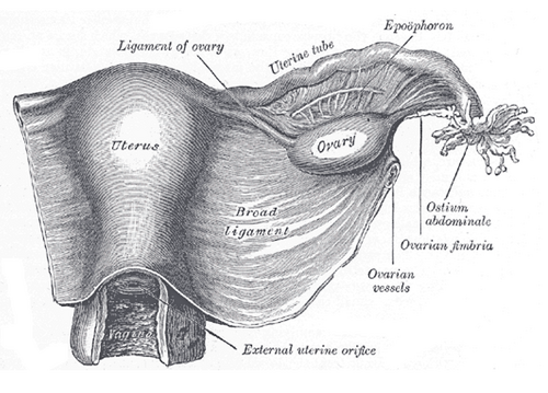
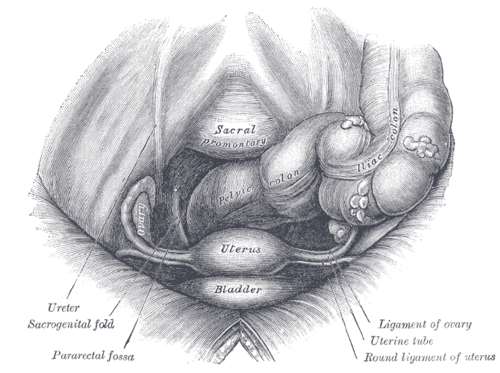
In the ovarian fossa on the lateral pelvic wall, between the external and internal iliac vessels; the obturator nerve runs across the floor of the fossa. Attached to the back of the broad ligament by the mesovarium.
- Parts
- Suspensory ligament of the ovary (infundibulopelvic ligament): fold of peritoneum from the ovary to the pelvic wall carrying the ovarian vessels, nerves and lymphatics
- Ligament of the ovary (utero-ovarian ligament): fibromuscular cord from the ovary to the uterus just below the tube (a remnant of the gubernaculum, continuous with the round ligament)
- Surface: covered by a modified cuboidal epithelium (not peritoneum), over the tunica albuginea, cortex (follicles) and medulla (vessels)
- Relations
The uterine tube arches over it, its fimbriae touching the ovary. The ureter lies just behind the ovarian fossa and crosses the pelvic brim close to the ovarian vessels in the suspensory ligament.
- Arterial supply
Ovarian artery from the abdominal aorta (at about L2), descending in the suspensory ligament; anastomoses with the ovarian branch of the uterine artery.
- Venous drainage
Pampiniform plexus → ovarian vein. Right ovarian vein → inferior vena cava; left ovarian vein → left renal vein.
- Lymphatic drainage
Lumbar (para-aortic) nodes, following the ovarian vessels (see pelvic and inguinal lymph drainage).
- Innervation
Ovarian plexus (from the aortic and renal plexuses) with the ovarian vessels; visceral pain is referred to the periumbilical region and lower abdomen (about T10–T11) verify


Clinical correlations
Ovarian torsion PANCE
- Mechanism / lesion
Twisting of the ovary (often with the tube) on its suspensory and ovarian ligaments, cutting off venous and then arterial flow. Usually an enlarged ovary (cyst or mass, often over 5 cm); also in pregnancy or with ovarian stimulation.
- Classic presentation
Sudden, severe unilateral pelvic pain with nausea and vomiting; tender adnexal mass. Doppler may still show flow (dual supply), so normal flow does not exclude torsion.
- Notes
A surgical emergency — laparoscopic detorsion, preserving the ovary whenever possible.
Ovarian cancer PANCE
- Mechanism / lesion
Most are epithelial (high-grade serous, many originating in the fimbriae). Risk factors — BRCA1/2, Lynch syndrome, family history, nulliparity, increasing age; oral contraceptives reduce risk.
- Classic presentation
Vague symptoms: bloating, early satiety, pelvic pain, urinary frequency; later ascites and an adnexal mass. Often diagnosed at an advanced stage. CA-125 helps evaluate a pelvic mass in postmenopausal women; there is no screening for average-risk women.
Functional ovarian cysts and cyst rupture
- Mechanism / lesion
Follicular or corpus luteum cysts; a ruptured corpus luteum can cause hemoperitoneum.
- Classic presentation
Sudden pelvic pain, often mid-cycle or luteal phase, sometimes after intercourse; free fluid on ultrasound. Most simple cysts resolve spontaneously.
Polycystic ovary syndrome PANCE
- Mechanism / lesion
Hyperandrogenism, ovulatory dysfunction and polycystic ovarian morphology (Rotterdam criteria — two of three).
- Classic presentation
Oligomenorrhea, hirsutism, acne, obesity and insulin resistance; increased risk of endometrial hyperplasia.
High-yield PANCE pearls
- Sudden unilateral pelvic pain + vomiting + adnexal mass → ovarian torsion; Doppler flow does not rule it out.
- Ovarian lymph → para-aortic nodes; left ovarian vein → left renal vein.
- Ovarian cancer: vague bloating/early satiety; BRCA; no screening for average risk.
- Obturator nerve in the ovarian fossa → ovarian disease may refer pain to the medial thigh.
Imaging anatomy
- CT
Staging of ovarian cancer (peritoneal implants, omental "cake", ascites, nodes).
- MRI
Characterizes indeterminate adnexal masses (e.g. dermoid fat, endometrioma blood).
- Ultrasound
Transvaginal ultrasound is first-line for adnexal masses (simple vs complex, solid parts, septations) and torsion (enlarged edematous ovary with peripheral follicles, "whirlpool sign" of the twisted pedicle). Risk is stratified with O-RADS.
- Key views & landmarks
- Transvaginal ultrasound with color Doppler
Physical exam
- Surface landmarks
The ovaries lie on the lateral pelvic walls on either side of the uterus; they are usually palpable (as small, mobile, slightly tender structures) only in thin premenopausal women.
- Palpation
Bimanual adnexal exam — size, mobility and tenderness. A palpable ovary in a postmenopausal woman needs evaluation, because the ovaries normally atrophy after menopause. verify
- Strength / function testing
Pregnancy test in all women of reproductive age with pelvic pain.
Self-quiz
A 24-year-old woman has sudden-onset severe right pelvic pain with vomiting that began while exercising. She has a tender right adnexal mass. Ultrasound shows a 7-cm enlarged right ovary with preserved arterial Doppler flow. What is the most appropriate next step?
Correct answer: C
The presentation is classic for ovarian torsion. Because the ovary has a dual blood supply (ovarian and uterine arteries), Doppler flow may be preserved. Clinical suspicion warrants urgent laparoscopy for detorsion.
Show answer
C
The presentation is classic for ovarian torsion. Because the ovary has a dual blood supply (ovarian and uterine arteries), Doppler flow may be preserved. Clinical suspicion warrants urgent laparoscopy for detorsion.
The left ovarian vein drains into which vessel?
Correct answer: B
Like the testicular veins: the left gonadal vein drains into the left renal vein, and the right drains directly into the IVC.
Show answer
B
Like the testicular veins: the left gonadal vein drains into the left renal vein, and the right drains directly into the IVC.
Ovarian cancer metastasizes first to which lymph nodes?
Correct answer: C
Ovarian lymph follows the ovarian vessels in the suspensory ligament to the lumbar (para-aortic) nodes, reflecting the gonad's embryologic origin high on the posterior abdominal wall.
Show answer
C
Ovarian lymph follows the ovarian vessels in the suspensory ligament to the lumbar (para-aortic) nodes, reflecting the gonad's embryologic origin high on the posterior abdominal wall.
Ovarian disease can refer pain to the medial thigh because which nerve runs across the floor of the ovarian fossa?
Correct answer: B
The ovary sits in the ovarian fossa on the lateral pelvic wall, and the obturator nerve runs across the floor of the fossa. The obturator nerve supplies skin of the medial thigh, so ovarian disease may refer pain there.
Show answer
B
The ovary sits in the ovarian fossa on the lateral pelvic wall, and the obturator nerve runs across the floor of the fossa. The obturator nerve supplies skin of the medial thigh, so ovarian disease may refer pain there.
Which structure carries the ovarian artery and vein to the ovary?
Correct answer: C
The suspensory ligament of the ovary (infundibulopelvic ligament) is a peritoneal fold from the ovary to the pelvic wall carrying the ovarian vessels, nerves and lymphatics. The ureter crosses the pelvic brim close to these vessels. The ligament of the ovary (utero-ovarian ligament) runs from the ovary to the uterus.
Show answer
C
The suspensory ligament of the ovary (infundibulopelvic ligament) is a peritoneal fold from the ovary to the pelvic wall carrying the ovarian vessels, nerves and lymphatics. The ureter crosses the pelvic brim close to these vessels. The ligament of the ovary (utero-ovarian ligament) runs from the ovary to the uterus.
Flashcards
See also
- Uterine tubes
- Uterus
- Broad ligament
- Lymph drainage of the pelvis and perineum (comparison table)