Brachial plexus
Network formed by the ventral rami of C5–T1 that supplies the motor and most of the sensory innervation of the upper limb.
Core anatomy
- Location
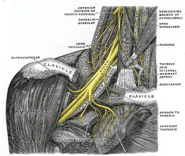
Extends from the neck into the axilla. The roots and trunks lie in the lateral cervical region (posterior triangle), the divisions lie posterior to the clavicle, and the cords lie in the axilla.
- Root values
Ventral (anterior) rami of C5, C6, C7, C8 and T1. A "prefixed" plexus has a large C4 contribution and a "postfixed" plexus a large T2 contribution.
- Origin
Ventral rami of spinal nerves C5–T1.
- Course
Roots emerge between the anterior and middle scalene muscles together with the subclavian artery. They unite to form three trunks: superior (C5–C6), middle (C7) and inferior (C8–T1). Each trunk splits behind the clavicle into an anterior and a posterior division. The anterior divisions of the superior and middle trunks form the lateral cord; the anterior division of the inferior trunk forms the medial cord; all three posterior divisions form the posterior cord. The cords are named by their position relative to the second part of the axillary artery, deep to pectoralis minor. Anterior divisions supply flexor (anterior) compartments; posterior divisions supply extensor (posterior) compartments.
- Branches
- From roots: dorsal scapular nerve (C5); long thoracic nerve (C5–C7)
- From superior trunk: suprascapular nerve (C5–C6); nerve to subclavius (C5–C6)
- From lateral cord: lateral pectoral nerve; musculocutaneous nerve; lateral root of the median nerve
- From medial cord: medial pectoral nerve; medial cutaneous nerves of the arm and forearm; ulnar nerve; medial root of the median nerve
- From posterior cord: upper and lower subscapular nerves; thoracodorsal nerve (C6–C8); axillary nerve; radial nerve
- Motor
All muscles of the upper limb except trapezius (spinal accessory nerve, CN XI).
- Sensory
Nearly all skin of the upper limb. Exceptions: the skin over the top of the shoulder (supraclavicular nerves, C3–C4, from the cervical plexus) and part of the medial arm near the axilla (intercostobrachial nerve, T2).
- Relations
Roots and trunks accompany the subclavian artery between the scalenes, while the subclavian vein passes anterior to anterior scalene. In the axilla the cords and their branches surround the axillary artery within the axillary sheath.
Muscles innervated
- Latissimus dorsi — Thoracodorsal nerve (C6, C7, C8), from the posterior cord of the brachial plexus.
- Rhomboid major and minor — Dorsal scapular nerve (C5, often with C4), from the C5 root of the brachial plexus.
Muscles innervated via branches
- Deltoid — via Axillary nerve
- Teres minor — via Axillary nerve
- Serratus anterior — via Long thoracic nerve
- Pronator teres — via Median nerve
- Biceps brachii — via Musculocutaneous nerve
- Triceps brachii — via Radial nerve
Branches
Found in
Mentioned in
Generated automatically from links in other entries.
Swipe sideways to see the whole diagram →

Clinical correlations
Upper plexus injury (Erb–Duchenne palsy) PANCE
- Mechanism / lesion
Injury to the C5–C6 roots or superior trunk from forcible widening of the neck–shoulder angle, e.g. shoulder dystocia during delivery or a fall onto the shoulder (motorcycle, contact sports).
- Classic presentation
"Waiter's tip" posture: arm adducted and medially rotated, elbow extended, forearm pronated. Loss of abduction (deltoid, supraspinatus), lateral rotation (infraspinatus, teres minor), elbow flexion and supination (biceps). Sensory loss over the lateral arm. Biceps reflex reduced or absent. In a newborn the Moro reflex is asymmetric.
Lower plexus injury (Klumpke palsy) PANCE
- Mechanism / lesion
Injury to the C8–T1 roots or inferior trunk from excessive upward traction on the arm, e.g. grabbing a support to break a fall, or delivery with the arm abducted overhead.
- Classic presentation
Weakness of the intrinsic hand muscles producing a claw hand, with sensory loss along the medial forearm and hand. If the T1 root is avulsed near the spinal cord, sympathetic fibres to the head are interrupted, causing ipsilateral Horner syndrome (ptosis, miosis, anhidrosis).
Pancoast (superior sulcus) tumor PANCE
- Mechanism / lesion
Apical lung carcinoma invading the inferior trunk (C8–T1) and the cervical sympathetic chain/stellate ganglion.
- Classic presentation
Shoulder pain radiating down the medial arm and forearm, wasting of the intrinsic hand muscles, and ipsilateral Horner syndrome, typically in a patient with a smoking history.
Thoracic outlet syndrome (neurogenic)
- Mechanism / lesion
Compression of the lower plexus (usually C8–T1/inferior trunk) in the interscalene triangle or costoclavicular space, e.g. by a cervical rib, fibrous band or scalene hypertrophy. The subclavian vessels may be compressed too (vascular TOS).
- Classic presentation
Pain and paresthesias along the medial forearm and hand, worse with overhead activity; weakness or wasting of the intrinsic hand muscles in advanced cases.
High-yield PANCE pearls
- Erb palsy = C5–C6, waiter's-tip posture; classic cause is shoulder dystocia.
- Klumpke palsy = C8–T1, claw hand ± Horner syndrome.
- Apical lung mass + Horner syndrome + ulnar-side arm pain → Pancoast tumor.
- Cords are named for their relation to the second part of the axillary artery.
- Mnemonic for the order: Roots, Trunks, Divisions, Cords, Branches.
Imaging anatomy
- X-ray
The plexus itself is not visible. Look for causes or associated injuries: a cervical rib or elongated C7 transverse process (thoracic outlet syndrome), fractures of the clavicle, first rib or cervical spine, and an apical lung mass (Pancoast tumor) on chest X-ray.
- CT
CT chest characterizes an apical (superior sulcus) mass. CT myelography can show pseudomeningoceles in traumatic root avulsion.
- MRI
MRI (including MR neurography) is the main modality for the plexus. It shows nerve thickening and T2/STIR hyperintensity after trauma or inflammation, pseudomeningoceles in root avulsion, tumor invasion, and denervation edema in the affected muscles.
- Ultrasound
Used for regional anesthesia. In the interscalene groove the C5–C7 roots/trunks appear as a vertical stack of round hypoechoic structures between the anterior and middle scalenes. In the supraclavicular region the divisions appear as a cluster of hypoechoic nodules lateral to the subclavian artery, above the first rib and pleura.
- Key views & landmarks
- Chest X-ray (PA) with attention to the lung apices
- Cervical spine AP to look for a cervical rib
- MRI brachial plexus protocol (coronal and sagittal STIR/T2)
Physical exam
- Surface landmarks
Roots and trunks: posterior triangle of the neck, deep to the lateral border of sternocleidomastoid. Cords: deep to pectoralis minor, inferomedial to the coracoid process.
- Strength / function testing
Localize by root using myotomes (ASIA key muscles): C5 elbow flexion; C6 wrist extension; C7 elbow extension; C8 finger flexion (FDP of the middle finger); T1 little-finger abduction. Reflexes: biceps (C5–C6), brachioradialis (C6), triceps (C7). Dermatomes: see the diagram.
Roos test (elevated arm stress test)
- Technique
Arms abducted to 90° and externally rotated with elbows flexed to 90°. The patient slowly opens and closes the hands for up to 3 minutes.
- Positive test
Reproduction of the patient's symptoms (pain, paresthesias, heaviness), or being unable to continue.
- Significance
Supports thoracic outlet syndrome. Specificity is limited.
Adson test
- Technique
The examiner palpates the radial pulse with the arm slightly abducted and extended. The patient extends the neck, rotates the head toward the tested side and takes a deep breath. verify
- Positive test
Diminished or absent radial pulse with reproduction of symptoms.
- Significance
Suggests vascular thoracic outlet compression. High false-positive rate in healthy people.
Self-quiz
A newborn delivered after a difficult vaginal delivery complicated by shoulder dystocia holds the right arm adducted and internally rotated, with the elbow extended and the forearm pronated. The Moro reflex is absent on the right. Injury to which spinal nerve roots is most likely?
Correct answer: A
This is the "waiter's tip" posture of Erb–Duchenne palsy (upper trunk, C5–C6). Deltoid and supraspinatus are weak (arm hangs adducted), infraspinatus and teres minor are weak (unopposed medial rotation), and biceps is weak (elbow extended, forearm pronated). C8–T1 (Klumpke) would produce a claw hand. Complete C5–T1 injury produces a flail arm.
Show answer
A
This is the "waiter's tip" posture of Erb–Duchenne palsy (upper trunk, C5–C6). Deltoid and supraspinatus are weak (arm hangs adducted), infraspinatus and teres minor are weak (unopposed medial rotation), and biceps is weak (elbow extended, forearm pronated). C8–T1 (Klumpke) would produce a claw hand. Complete C5–T1 injury produces a flail arm.
A 61-year-old man with a 50-pack-year smoking history has 3 months of right shoulder pain radiating to the medial forearm. Exam shows wasting of the right interossei, right ptosis and a small right pupil. Which part of the brachial plexus is most likely involved?
Correct answer: D
A Pancoast tumor at the lung apex invades the inferior trunk (C8–T1), producing medial arm and forearm pain and intrinsic hand weakness. Involvement of the adjacent cervical sympathetic chain causes Horner syndrome. The superior trunk (C5–C6) would affect shoulder abduction and elbow flexion instead.
Show answer
D
A Pancoast tumor at the lung apex invades the inferior trunk (C8–T1), producing medial arm and forearm pain and intrinsic hand weakness. Involvement of the adjacent cervical sympathetic chain causes Horner syndrome. The superior trunk (C5–C6) would affect shoulder abduction and elbow flexion instead.
Which nerve arises from the posterior cord of the brachial plexus?
Correct answer: D
The posterior cord gives the upper and lower subscapular, thoracodorsal, axillary and radial nerves. The lateral pectoral and musculocutaneous nerves come from the lateral cord, the suprascapular nerve from the superior trunk, and the ulnar nerve from the medial cord.
Show answer
D
The posterior cord gives the upper and lower subscapular, thoracodorsal, axillary and radial nerves. The lateral pectoral and musculocutaneous nerves come from the lateral cord, the suprascapular nerve from the superior trunk, and the ulnar nerve from the medial cord.
The cords of the brachial plexus are named according to their relationship to which structure?
Correct answer: C
The lateral, medial and posterior cords are named for their position around the second part of the axillary artery, which lies posterior to pectoralis minor.
Show answer
C
The lateral, medial and posterior cords are named for their position around the second part of the axillary artery, which lies posterior to pectoralis minor.
Which nerve arises directly from the roots of the brachial plexus?
Correct answer: C
The long thoracic nerve (C5–C7) and the dorsal scapular nerve (C5) arise from the roots. The suprascapular nerve comes from the superior trunk, the thoracodorsal from the posterior cord, and the lateral pectoral and musculocutaneous nerves from the lateral cord.
Show answer
C
The long thoracic nerve (C5–C7) and the dorsal scapular nerve (C5) arise from the roots. The suprascapular nerve comes from the superior trunk, the thoracodorsal from the posterior cord, and the lateral pectoral and musculocutaneous nerves from the lateral cord.
Flashcards
See also
- Axilla
- Axillary artery
- Upper limb nerve injuries (comparison table)
- A drooping eyelid and shoulder pain in a smoker (clinical case)