Carpal tunnel
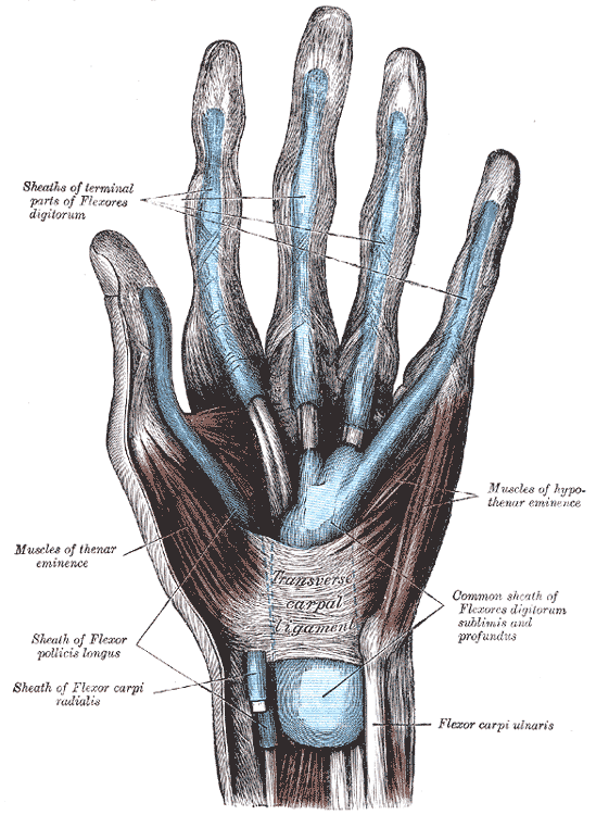
Osseofibrous tunnel on the palmar wrist, formed by the carpal bones and the flexor retinaculum, that carries the median nerve and nine long flexor tendons.
Core anatomy
- Location
Palmar aspect of the wrist, deep to the flexor retinaculum.
- Boundaries
- roof
- Flexor retinaculum (transverse carpal ligament). Medial attachments: pisiform and hook of hamate. Lateral attachments: scaphoid tubercle and trapezium tubercle.
- floor and walls
- The concave palmar arch formed by the carpal bones
- Contents
- Median nerve (the most superficial structure in the tunnel)
- Four tendons of flexor digitorum superficialis
- Four tendons of flexor digitorum profundus
- One tendon of flexor pollicis longus
- Synovial sheaths: the common flexor sheath (ulnar bursa) for FDS/FDP and a separate sheath (radial bursa) for FPL
- Communications
Leads from the anterior compartment of the forearm to the central compartment of the palm.
- Notes
Not in the tunnel: the ulnar nerve and artery (superficial to the retinaculum, in Guyon's canal); the palmar cutaneous branch of the median nerve and the palmaris longus tendon (superficial to the retinaculum). The flexor carpi radialis tendon runs in its own split within the retinaculum (a separate compartment). Mnemonic: 9 tendons + 1 nerve.
Swipe sideways to see the whole diagram →

Clinical correlations
Carpal tunnel syndrome PANCE
- Mechanism / lesion
Anything that reduces the space or enlarges the contents compresses the median nerve: pregnancy (fluid), hypothyroidism, diabetes, rheumatoid arthritis (flexor tenosynovitis), obesity, acromegaly, dialysis-related amyloidosis, distal radius fracture, lunate dislocation, and repetitive wrist use.
- Classic presentation
Nocturnal paresthesias of the thumb, index, middle and radial half of the ring finger. Hand-shaking relief. Clumsiness. Late thenar atrophy and weakness of thumb abduction. Thenar skin sensation is spared.
Carpal tunnel release
- Mechanism / lesion
Surgical division of the flexor retinaculum (open or endoscopic) for severe or refractory symptoms.
- Classic presentation
The recurrent (thenar motor) branch of the median nerve is at risk, particularly with anatomic variants where it passes through the retinaculum. Injury leaves the patient unable to oppose the thumb.
High-yield PANCE pearls
- Carpal tunnel contents: median nerve + 9 flexor tendons (4 FDS, 4 FDP, 1 FPL).
- The ulnar nerve and artery are not in the carpal tunnel (Guyon's canal).
- CTS associations: pregnancy, hypothyroidism, diabetes, RA, acromegaly, amyloidosis.
- Diagnosis is clinical (Tinel, Phalen, Durkan); nerve conduction studies/EMG confirm and grade severity.
Imaging anatomy
- X-ray
The carpal tunnel view (axial projection with the wrist dorsiflexed) shows the pisiform, the hook of hamate, and the tubercles of the scaphoid and trapezium, the attachments of the retinaculum. It is useful for hook of hamate fractures and bony encroachment.
- CT
Hook of hamate fractures and bony masses.
- MRI
Swelling of the median nerve proximally, flattening distally, increased T2 signal, bowing of the flexor retinaculum, and tenosynovitis or masses.
- Ultrasound
Shows the median nerve's cross-sectional area at the tunnel inlet, flexor tenosynovitis, anatomic variants (bifid median nerve, persistent median artery), and ganglia. Guides corticosteroid injection.
- Key views & landmarks
- Carpal tunnel (axial) radiograph
- Ultrasound transverse at the pisiform and hook of hamate levels
Physical exam
- Surface landmarks
The distal wrist crease roughly marks the proximal edge of the flexor retinaculum, which extends distally into the palm. Palpable attachments: pisiform (base of the hypothenar eminence), hook of hamate (deep, distal and radial to the pisiform), scaphoid tubercle and trapezium tubercle (base of the thenar eminence).
- Palpation
Palpate the four retinacular attachments. Look for thenar atrophy (flattening of the lateral eminence).
- Strength / function testing
Thumb abduction strength (APB). Two-point discrimination on the index pad. Thenar sensation (spared in CTS).
Tinel sign
- Technique
Tap over the carpal tunnel.
- Positive test
Tingling in the median-innervated digits.
- Significance
Carpal tunnel syndrome.
Phalen test
- Technique
Full wrist flexion for 60 seconds.
- Positive test
Median-distribution paresthesias.
- Significance
Carpal tunnel syndrome.
Carpal compression (Durkan) test
- Technique
Direct pressure over the carpal tunnel for up to 30 seconds.
- Positive test
Median-distribution paresthesias.
- Significance
Carpal tunnel syndrome.
Self-quiz
Which structure passes through the carpal tunnel?
Correct answer: C
The carpal tunnel contains the median nerve and nine tendons: 4 FDS, 4 FDP and FPL. The ulnar nerve and artery pass through Guyon's canal. The palmar cutaneous branch and palmaris longus pass superficial to the retinaculum.
Show answer
C
The carpal tunnel contains the median nerve and nine tendons: 4 FDS, 4 FDP and FPL. The ulnar nerve and artery pass through Guyon's canal. The palmar cutaneous branch and palmaris longus pass superficial to the retinaculum.
A 48-year-old woman with fatigue, cold intolerance and weight gain reports bilateral nocturnal hand numbness in the thumb, index and middle fingers. Which test is most appropriate to evaluate an underlying cause?
Correct answer: B
Her symptoms suggest hypothyroidism, a recognized cause of carpal tunnel syndrome (myxedematous tissue deposition). Check TSH. Other associations include pregnancy, diabetes, RA and acromegaly.
Show answer
B
Her symptoms suggest hypothyroidism, a recognized cause of carpal tunnel syndrome (myxedematous tissue deposition). Check TSH. Other associations include pregnancy, diabetes, RA and acromegaly.
The medial attachments of the flexor retinaculum are which structures?
Correct answer: B
The flexor retinaculum attaches medially to the pisiform and hook of hamate and laterally to the scaphoid tubercle and trapezium.
Show answer
B
The flexor retinaculum attaches medially to the pisiform and hook of hamate and laterally to the scaphoid tubercle and trapezium.
During carpal tunnel release, injury to which branch would cause loss of thumb opposition?
Correct answer: B
The recurrent branch supplies abductor pollicis brevis, opponens pollicis and the superficial head of FPB. Injuring it causes loss of opposition. The palmar cutaneous branch is purely sensory.
Show answer
B
The recurrent branch supplies abductor pollicis brevis, opponens pollicis and the superficial head of FPB. Injuring it causes loss of opposition. The palmar cutaneous branch is purely sensory.
In carpal tunnel syndrome, sensation over the thenar eminence is usually normal. Why?
Correct answer: D
The palmar cutaneous branch arises proximal to the flexor retinaculum and passes superficial to it, so it isn't compressed in the tunnel. Reduced thenar sensation points to a more proximal lesion (e.g. pronator syndrome). The recurrent branch is motor to the thenar muscles.
Show answer
D
The palmar cutaneous branch arises proximal to the flexor retinaculum and passes superficial to it, so it isn't compressed in the tunnel. Reduced thenar sensation points to a more proximal lesion (e.g. pronator syndrome). The recurrent branch is motor to the thenar muscles.
Flashcards
See also
- Median nerve
- Wrist joint
- Numb fingers that wake her at night (clinical case)