Ulnar nerve
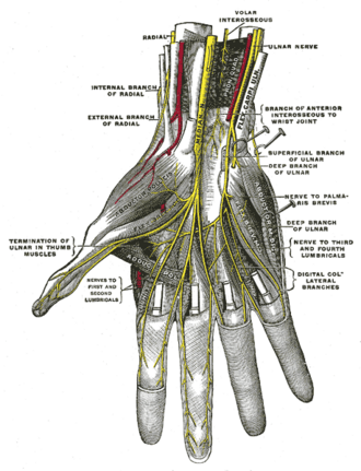
Medial cord branch that passes behind the medial epicondyle and through Guyon's canal. It supplies most intrinsic hand muscles and the medial 1½ digits.
Core anatomy
- Location
Medial arm → posterior to the medial epicondyle (cubital tunnel) → medial anterior forearm → Guyon's canal → hand.
- Root values
C8, T1 (often C7)
- Origin
Terminal branch of the medial cord of the brachial plexus.
- Course
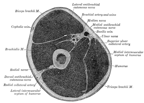
Descends medial to the brachial artery in the arm. At mid-arm it pierces the medial intermuscular septum with the superior ulnar collateral artery and descends on the medial head of triceps. It passes posterior to the medial epicondyle of the humerus in the ulnar groove/cubital tunnel, where it is subcutaneous and easily compressed (the "funny bone"). It enters the forearm between the two heads of flexor carpi ulnaris (FCU) and runs between FCU and FDP, with the ulnar artery on its lateral side in the distal forearm. The dorsal cutaneous branch arises in the distal forearm. The nerve enters the hand superficial to the flexor retinaculum through Guyon's canal (the ulnar canal, between the pisiform and the hook of hamate), where it divides into superficial (sensory) and deep (motor) branches.
- Branches
- Muscular branches to flexor carpi ulnaris and the medial half of flexor digitorum profundus (digits 4–5)
- Palmar cutaneous branch: medial palm
- Dorsal cutaneous branch: medial dorsum of the hand and dorsal medial 1½ digits (arises before Guyon's canal)
- Superficial branch: palmaris brevis; palmar skin of the medial 1½ digits
- Deep branch: hypothenar muscles, medial two lumbricals, all interossei, adductor pollicis, deep head of flexor pollicis brevis
- Motor
Forearm: FCU and the medial half of FDP. Hand: most intrinsic muscles, including abductor digiti minimi, flexor digiti minimi brevis, opponens digiti minimi, the 3rd and 4th lumbricals, all palmar and dorsal interossei, adductor pollicis, the deep head of flexor pollicis brevis, and palmaris brevis.
- Sensory
Palmar and dorsal skin of the medial 1½ digits (little finger and medial half of the ring finger) and the corresponding medial palm and dorsum. Most reliable test site: the pulp of the little finger.
Other structures innervated
Mentioned in
Generated automatically from links in other entries.
Swipe sideways to see the whole diagram →


Clinical correlations
Cubital tunnel syndrome PANCE
- Mechanism / lesion
Compression or traction at the elbow: prolonged elbow flexion, leaning on the elbows, nerve subluxation, cubitus valgus after an old fracture, or fracture of the medial epicondyle. It is the second most common upper-limb entrapment neuropathy after carpal tunnel syndrome.
- Classic presentation
Paresthesias in the little finger and the medial half of the ring finger, including the dorsal ulnar hand (the dorsal cutaneous branch arises proximal to the wrist). Medial elbow pain and a positive Tinel sign at the elbow. Weak grip, pinch and finger abduction; interosseous wasting in advanced cases.
Guyon's canal (ulnar tunnel) syndrome PANCE
- Mechanism / lesion
Compression at the wrist, e.g. cyclists leaning on handlebars ("handlebar palsy"), fracture of the hook of hamate (golf, baseball, racquet sports), ganglion cyst, or ulnar artery aneurysm/thrombosis.
- Classic presentation
Intrinsic hand weakness ± palmar sensory loss in the medial 1½ digits, with sparing of dorsal hand sensation. FCU and FDP are spared.
Ulnar claw hand PANCE
- Mechanism / lesion
Loss of the medial lumbricals and interossei with intact long finger extensors and flexors.
- Classic presentation
When the patient tries to extend the fingers, the 4th and 5th digits hyperextend at the MCP and flex at the IP joints. Clawing is worse with distal (wrist) lesions than proximal (elbow) lesions, because a proximal lesion also paralyzes the medial FDP (the "ulnar paradox").
High-yield PANCE pearls
- Hook of hamate fracture or cycling → ulnar nerve compression at Guyon's canal; dorsal hand sensation spared.
- Froment sign = weak adductor pollicis (thumb IP flexes via FPL to compensate).
- Ulnar claw: 4th and 5th digits clawed; the ulnar paradox makes distal lesions claw more.
- Elbow (cubital tunnel) lesion: dorsal ulnar hand numbness + FCU/FDP weakness in addition to intrinsic weakness.
Imaging anatomy
- X-ray
Not visible. Elbow films show a medial epicondyle fracture, osteophytes or valgus deformity. A carpal tunnel view (or CT) shows a hook of hamate fracture, which is often missed on routine wrist views.
- CT
CT is sensitive for hook of hamate fractures.
- MRI
Increased T2 signal and enlargement of the nerve in the cubital tunnel. Denervation edema of the intrinsic hand muscles. Masses in Guyon's canal.
- Ultrasound
Measures nerve enlargement at the medial epicondyle. Dynamic ultrasound during elbow flexion shows subluxation or dislocation of the nerve over the medial epicondyle. At the wrist, identifies ganglia and ulnar artery thrombosis or aneurysm in Guyon's canal.
- Key views & landmarks
- Elbow AP and lateral; carpal tunnel view for the hook of hamate
- Dynamic ultrasound at the cubital tunnel, elbow extended → flexed
Physical exam
- Surface landmarks
Palpable in the groove posterior to the medial epicondyle. At the wrist it lies lateral to the pisiform and medial to the ulnar artery, lateral to the FCU tendon.
- Palpation
Roll the nerve in the ulnar groove with the elbow partly flexed. Check for tenderness, thickening, or subluxation with flexion.
- Strength / function testing
Finger abduction against resistance (first dorsal interosseous). Adduction (card test for palmar interossei). FDP to the little finger (DIP flexion). Sensation at the pulp of the little finger, and on the dorsal ulnar hand to distinguish elbow from wrist lesions.
Froment sign
- Technique
The patient holds a sheet of paper between the thumb and the radial side of the index finger (key pinch) while the examiner pulls it away.
- Positive test
The thumb IP joint flexes (FPL, median nerve) to compensate for weak adductor pollicis.
- Significance
Ulnar nerve palsy.
Wartenberg sign
- Technique
Ask the patient to hold all fingers extended and adducted.
- Positive test
The little finger drifts into abduction and can't be adducted.
- Significance
Weakness of the third palmar interosseous (ulnar nerve), with unopposed pull of extensor digiti minimi.
Elbow flexion test
- Technique
The patient holds the elbow fully flexed with the wrist extended for about 1 minute.
- Positive test
Paresthesias in the ulnar distribution.
- Significance
Suggests cubital tunnel syndrome.
Tinel sign (elbow)
- Technique
Tap over the nerve in the ulnar groove behind the medial epicondyle.
- Positive test
Tingling radiating to the little and ring fingers.
- Significance
Suggests ulnar neuropathy at the elbow.
Self-quiz
A 38-year-old competitive cyclist has numbness of the palmar little finger and weakness of finger abduction after long rides. Sensation over the dorsal ulnar hand is normal, and flexion of the little finger DIP joint is strong. Where is the nerve compressed?
Correct answer: C
Sparing of the dorsal cutaneous branch (dorsal hand sensation) and of FDP localizes the lesion distal to the forearm, at Guyon's canal. The median nerve, not the ulnar, runs in the carpal tunnel.
Show answer
C
Sparing of the dorsal cutaneous branch (dorsal hand sensation) and of FDP localizes the lesion distal to the forearm, at Guyon's canal. The median nerve, not the ulnar, runs in the carpal tunnel.
When a patient pinches a piece of paper between the thumb and index finger, the thumb IP joint flexes. Weakness of which muscle does this compensate for?
Correct answer: B
This is the Froment sign. The patient recruits FPL (median nerve) to compensate for a weak adductor pollicis (deep branch of the ulnar nerve).
Show answer
B
This is the Froment sign. The patient recruits FPL (median nerve) to compensate for a weak adductor pollicis (deep branch of the ulnar nerve).
Compared with an ulnar nerve lesion at the wrist, why does a lesion at the elbow typically produce less clawing of the 4th and 5th digits?
Correct answer: B
A proximal lesion paralyzes the medial FDP, so there is less unopposed flexion at the IP joints. That's the ulnar paradox: the more proximal lesion produces less clawing.
Show answer
B
A proximal lesion paralyzes the medial FDP, so there is less unopposed flexion at the IP joints. That's the ulnar paradox: the more proximal lesion produces less clawing.
The ulnar nerve passes into the forearm between the two heads of which muscle?
Correct answer: C
The ulnar nerve enters the forearm between the humeral and ulnar heads of flexor carpi ulnaris. The median nerve passes between the heads of pronator teres.
Show answer
C
The ulnar nerve enters the forearm between the humeral and ulnar heads of flexor carpi ulnaris. The median nerve passes between the heads of pronator teres.
A patient with paresthesias of the little finger also has numbness over the dorsal ulnar side of the hand. Where is the ulnar nerve lesion most likely?
Correct answer: C
The dorsal cutaneous branch arises in the distal forearm, before Guyon's canal. Dorsal ulnar hand numbness therefore places the lesion proximal to that branch, classically the cubital tunnel. Guyon's canal lesions spare dorsal sensation.
Show answer
C
The dorsal cutaneous branch arises in the distal forearm, before Guyon's canal. Dorsal ulnar hand numbness therefore places the lesion proximal to that branch, classically the cubital tunnel. Guyon's canal lesions spare dorsal sensation.
Flashcards
See also
- Elbow joint
- Wrist joint
- Upper limb nerve injuries (comparison table)
- A drooping eyelid and shoulder pain in a smoker (clinical case)