Esophagus
Muscular tube from the pharynx (C6) through the posterior mediastinum to the stomach (T11). Its constrictions, portosystemic anastomosis and relations explain many board classics.
Core anatomy
- Location
Neck → superior and posterior mediastinum → through the esophageal hiatus of the diaphragm → abdomen.
- Parts
- Begins at the inferior border of the cricoid cartilage (C6) as the continuation of the pharynx; ends at the cardiac orifice of the stomach (about T11). About 25 cm long
- Muscle: upper third skeletal, middle third mixed, lower third smooth
- Upper esophageal sphincter (cricopharyngeus) and a physiological lower esophageal sphincter at the GE junction
- Three constrictions (where foreign bodies lodge): cervical (cricopharyngeus, about 15 cm from the incisors), thoracic (crossed by the aortic arch and left main bronchus, about 25 cm), diaphragmatic (esophageal hiatus, about 40 cm). verify
- Relations
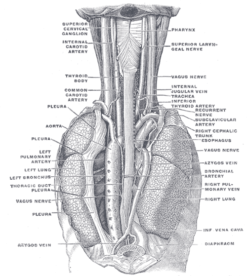
Posterior to the trachea and the left atrium (enlargement of the LA indents it). The thoracic aorta lies to its left above and then passes posterior to it. The thoracic duct and azygos vein lie posterior and to the right. The vagus nerves form the esophageal plexus on its surface, then the anterior (mostly left vagus) and posterior (mostly right vagus) vagal trunks pass with it through the hiatus at T10.
- Arterial supply
Cervical: inferior thyroid arteries. Thoracic: esophageal branches of the thoracic aorta (and bronchial arteries). Abdominal: left gastric artery (and left inferior phrenic).
- Venous drainage
Cervical → inferior thyroid veins. Thoracic → azygos and hemiazygos veins. Abdominal part → left gastric vein (portal system). The submucosal plexus of the distal esophagus is a portosystemic anastomosis.
- Lymphatic drainage
Deep cervical, posterior mediastinal, and left gastric/celiac nodes by region. Lymph drains longitudinally, so tumors spread widely.
- Innervation
Vagus nerves (esophageal plexus, recurrent laryngeal nerves to the cervical part) and sympathetic trunks.
Forms a boundary of
Found in
Mentioned in
- Azygos vein
- Heart
- Lungs
- Main bronchi
- Mitral valve
- Pharynx
- Thoracic aorta
- Thoracic duct
- Trachea
- Vagus nerve (CN X)
Generated automatically from links in other entries.

Clinical correlations
Esophageal varices PANCE
- Mechanism / lesion
Portal hypertension (usually cirrhosis) diverts portal blood through the left gastric vein → esophageal submucosal veins → azygos vein, dilating the distal esophageal veins.
- Classic presentation
Painless massive hematemesis in a patient with liver disease. Life-threatening; managed with resuscitation, octreotide, antibiotics and urgent endoscopic band ligation.
Boerhaave syndrome PANCE
- Mechanism / lesion
Full-thickness rupture, usually of the distal left posterolateral wall, from a sudden rise in pressure during forceful vomiting (often after binge drinking or eating).
- Classic presentation
Severe retrosternal/epigastric pain after vomiting, subcutaneous emphysema, and Hamman crunch (pneumomediastinum). Left pleural effusion. Mackler triad: vomiting, chest pain, subcutaneous emphysema.
Mallory–Weiss tear PANCE
- Mechanism / lesion
Mucosal (partial-thickness) laceration at the GE junction from retching or vomiting.
- Classic presentation
Hematemesis after repeated retching; usually self-limited.
Achalasia PANCE
- Mechanism / lesion
Loss of inhibitory neurons in the myenteric (Auerbach) plexus → failure of LES relaxation and absent peristalsis. Chagas disease is a secondary cause.
- Classic presentation
Progressive dysphagia to both solids and liquids, regurgitation, weight loss. Barium swallow shows a "bird's beak". Diagnosed with manometry.
Esophageal cancer PANCE
- Mechanism / lesion
Squamous cell carcinoma (upper/middle third; smoking, alcohol, hot beverages). Adenocarcinoma (distal third; GERD → Barrett esophagus, obesity).
- Classic presentation
Progressive dysphagia (solids → liquids), weight loss, odynophagia.
High-yield PANCE pearls
- Three constrictions: cricopharyngeus, aortic arch/left main bronchus, diaphragm → foreign body impaction sites.
- Boerhaave = full-thickness rupture (emergency); Mallory–Weiss = mucosal tear.
- Varices: left gastric (portal) ↔ esophageal/azygos (systemic).
- Achalasia: dysphagia to solids and liquids, bird's beak on barium, manometry for diagnosis.
- Barrett esophagus → adenocarcinoma (distal); smoking/alcohol → squamous cell (upper/middle).
Imaging anatomy
- X-ray
Swallowed coins in the esophagus appear en face (as a disk) on a frontal view (a coin in the trachea tends to appear on edge). Pneumomediastinum, left effusion and subcutaneous air in Boerhaave. An air–fluid level behind the heart can mean a hiatal hernia or achalasia.
- CT
CT (with oral water-soluble contrast) for suspected perforation: extraluminal air or contrast, mediastinal fluid. Cancer staging.
- Ultrasound
Endoscopic ultrasound (EUS) stages the depth of tumor invasion and nearby nodes.
- Key views & landmarks
- Barium esophagram: bird's beak (achalasia), corkscrew (diffuse spasm), strictures, rings, webs
- Water-soluble contrast study first when perforation is suspected (barium can cause mediastinitis)
- Endoscopy (EGD) for mucosal diagnosis, biopsy and variceal banding
Physical exam
- Surface landmarks
The cervical part lies behind the trachea, slightly to the left, where it is approached surgically.
- Palpation
Crepitus in the neck or chest wall (subcutaneous emphysema) with perforation.
- Strength / function testing
Auscultate for Hamman crunch (a crunching sound synchronous with the heartbeat) in pneumomediastinum.
Self-quiz
After a night of heavy drinking and repeated forceful vomiting, a 45-year-old man has severe chest pain and crepitus over the neck. CXR shows pneumomediastinum and a left pleural effusion. What is the diagnosis?
Correct answer: B
Boerhaave syndrome is a full-thickness esophageal rupture, usually distal left posterolateral, causing mediastinal air and a left effusion. It's a surgical emergency. A Mallory–Weiss tear is mucosal and presents with hematemesis.
Show answer
B
Boerhaave syndrome is a full-thickness esophageal rupture, usually distal left posterolateral, causing mediastinal air and a left effusion. It's a surgical emergency. A Mallory–Weiss tear is mucosal and presents with hematemesis.
Esophageal varices in portal hypertension form at an anastomosis between which veins?
Correct answer: B
The left gastric vein (portal) anastomoses with esophageal veins draining to the azygos (systemic) in the distal esophagus. The other options describe rectal and umbilical anastomoses.
Show answer
B
The left gastric vein (portal) anastomoses with esophageal veins draining to the azygos (systemic) in the distal esophagus. The other options describe rectal and umbilical anastomoses.
A child swallowed a coin. At which site is it most likely to lodge?
Correct answer: B
The cricopharyngeal (cervical) constriction is the narrowest point of the esophagus and the most common site of foreign body impaction in children. The other constrictions are at the aortic arch and the diaphragm.
Show answer
B
The cricopharyngeal (cervical) constriction is the narrowest point of the esophagus and the most common site of foreign body impaction in children. The other constrictions are at the aortic arch and the diaphragm.
A 40-year-old woman has 1 year of progressive dysphagia to both solids and liquids with regurgitation of undigested food. Barium swallow shows a dilated esophagus with a narrow distal "bird's beak." What confirms the diagnosis?
Correct answer: B
Achalasia is confirmed by esophageal manometry (incomplete LES relaxation, absent peristalsis). Endoscopy is done to exclude pseudoachalasia from a tumor at the GE junction.
Show answer
B
Achalasia is confirmed by esophageal manometry (incomplete LES relaxation, absent peristalsis). Endoscopy is done to exclude pseudoachalasia from a tumor at the GE junction.
Adenocarcinoma of the distal esophagus is most strongly associated with which condition?
Correct answer: D
Adenocarcinoma arises in the distal third, linked to GERD → Barrett esophagus and obesity. Squamous cell carcinoma affects the upper and middle thirds and is linked to smoking, alcohol and hot beverages.
Show answer
D
Adenocarcinoma arises in the distal third, linked to GERD → Barrett esophagus and obesity. Squamous cell carcinoma affects the upper and middle thirds and is linked to smoking, alcohol and hot beverages.