Basal ganglia
Deep gray-matter nuclei that modulate movement through direct and indirect pathways. Their disorders cause either too little movement (Parkinson disease) or too much (Huntington chorea, hemiballismus).
Core anatomy
- Location
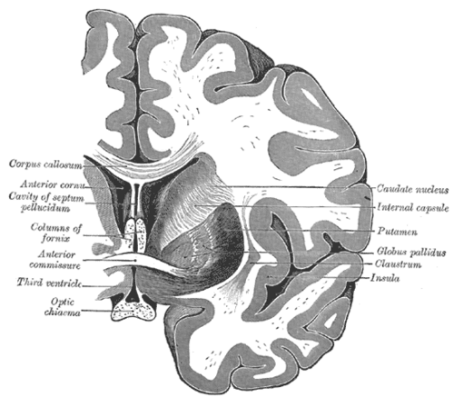
Deep in each cerebral hemisphere around the internal capsule and thalamus.
- Parts
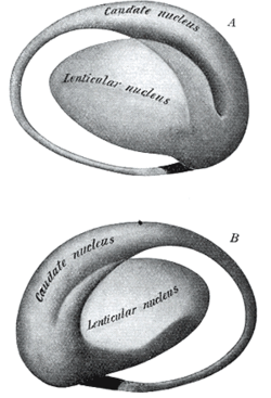
- Striatum = caudate nucleus (head bulges into the lateral ventricle) + putamen — the main input nucleus (from cortex and substantia nigra)
- Lentiform nucleus = putamen + globus pallidus
- Globus pallidus internus (GPi) — main output (inhibitory, GABA) to the thalamus; externus (GPe) — part of the indirect pathway
- Subthalamic nucleus (excitatory, glutamate → GPi)
- Substantia nigra in the midbrain — pars compacta (dopamine to the striatum) and pars reticulata (output)
- Direct pathway (cortex → striatum → GPi → thalamus) increases movement; indirect pathway (via GPe and subthalamic nucleus) decreases it. Dopamine excites the direct pathway (D1) and inhibits the indirect pathway (D2) — both favor movement.
- Relations
The internal capsule separates the caudate and thalamus (medially) from the lentiform nucleus (laterally).
- Arterial supply
Lenticulostriate arteries (from the MCA), recurrent artery of Heubner (ACA), anterior choroidal artery (ICA) — see cerebral arteries (ACA, MCA, PCA).
- Venous drainage
Deep cerebral veins → great cerebral vein → straight sinus.
- Notes
Loops with the cortex and thalamus; no peripheral nerves. Lesions produce contralateral movement disorders.


Clinical correlations
Parkinson disease PANCE
- Mechanism / lesion
Loss of dopamine from the substantia nigra → underactive direct pathway, overactive indirect pathway → too little movement.
- Classic presentation
Resting tremor, cogwheel rigidity, bradykinesia, postural instability, shuffling gait, masked face. Levodopa-carbidopa is most effective; dopamine agonists, MAO-B inhibitors. Drug-induced parkinsonism from D2 blockers (antipsychotics, metoclopramide).
Huntington disease PANCE
- Mechanism / lesion
Autosomal dominant CAG trinucleotide repeat in the HTT gene (chromosome 4) with anticipation; loss of GABAergic neurons in the caudate (and putamen).
- Classic presentation
Onset about 30–50 years: chorea, psychiatric changes (depression, irritability, suicide risk), and dementia. MRI: caudate atrophy with enlarged ("boxcar") lateral ventricles. Tetrabenazine for chorea; genetic counseling.
Hemiballismus PANCE
- Mechanism / lesion
Lesion (usually a lacunar stroke) of the contralateral subthalamic nucleus.
- Classic presentation
Sudden violent flinging movements of one arm and/or leg on the side opposite the lesion.
Hypertensive hemorrhage and Wilson disease
- Mechanism / lesion
Rupture of lenticulostriate arteries (Charcot–Bouchard microaneurysms); copper deposition in the lentiform nucleus.
- Classic presentation
Putamen is the most common site of hypertensive intracerebral hemorrhage (contralateral hemiparesis). Wilson disease: young patient with tremor ("wing-beating"), dystonia, psychiatric symptoms, liver disease and Kayser–Fleischer rings; low ceruloplasmin.
High-yield PANCE pearls
- Chorea + psychiatric symptoms + dementia + family history → Huntington (CAG repeats, caudate atrophy).
- Sudden flinging of one arm → contralateral subthalamic nucleus lesion.
- Parkinsonism in a patient on antipsychotics or metoclopramide → drug-induced (D2 blockade).
- Most common site of hypertensive ICH → putamen.
- Young patient + movement disorder + liver disease → Wilson disease (ceruloplasmin, KF rings).
Imaging anatomy
- CT
Hypertensive hemorrhage in the putamen.
- MRI
Caudate atrophy in Huntington disease; putaminal/basal ganglia T2 changes in Wilson disease; usually normal in Parkinson disease (used to exclude mimics).
- Key views & landmarks
- DaT-SPECT (dopamine transporter) reduced in Parkinson disease, normal in essential tremor and drug-induced parkinsonism
Physical exam
- Strength / function testing
Observe at rest and during distraction for tremor and chorea; tone (rigidity, cogwheeling), bradykinesia (finger taps, foot taps with decrement), gait (arm swing, turning), pull test for postural instability, handwriting (micrographia).
Self-quiz
A 40-year-old man has irritability, depression and brief, dance-like involuntary movements. His father had similar symptoms and died in his 50s. Which structure shows atrophy on MRI?
Correct answer: B
Huntington disease (autosomal dominant CAG repeat) causes caudate atrophy with chorea, psychiatric symptoms and dementia.
Show answer
B
Huntington disease (autosomal dominant CAG repeat) causes caudate atrophy with chorea, psychiatric symptoms and dementia.
A 70-year-old with hypertension suddenly develops wild flinging movements of the left arm. Where is the lesion?
Correct answer: B
Hemiballismus results from a lesion (usually lacunar infarct) of the contralateral subthalamic nucleus — here the right.
Show answer
B
Hemiballismus results from a lesion (usually lacunar infarct) of the contralateral subthalamic nucleus — here the right.
How does dopamine from the substantia nigra affect the direct and indirect pathways?
Correct answer: C
Dopamine excites the direct pathway (D1) and inhibits the indirect pathway (D2); both effects favor movement. Its loss in Parkinson disease leaves too little movement.
Show answer
C
Dopamine excites the direct pathway (D1) and inhibits the indirect pathway (D2); both effects favor movement. Its loss in Parkinson disease leaves too little movement.
A 19-year-old has a new tremor, personality change and abnormal liver tests. Slit-lamp exam shows brown rings at the corneal margin. Which lab finding is expected?
Correct answer: E
Wilson disease deposits copper in the lentiform nucleus and liver, causing tremor, dystonia, psychiatric symptoms and Kayser–Fleischer rings, with a low ceruloplasmin.
Show answer
E
Wilson disease deposits copper in the lentiform nucleus and liver, causing tremor, dystonia, psychiatric symptoms and Kayser–Fleischer rings, with a low ceruloplasmin.
What is the most common site of hypertensive intracerebral hemorrhage?
Correct answer: A
The putamen is the most common site, from rupture of lenticulostriate arteries (Charcot–Bouchard microaneurysms), followed by the thalamus.
Show answer
A
The putamen is the most common site, from rupture of lenticulostriate arteries (Charcot–Bouchard microaneurysms), followed by the thalamus.