Femur
The longest and strongest bone in the body. Femoral neck fractures in older adults, slipped capital femoral epiphysis and avascular necrosis of the head are its high-yield problems.
Core anatomy
- Location
The single bone of the thigh, from the acetabulum of the hip bone to the knee joint.
- Classification
Long bone
- Key features & landmarks
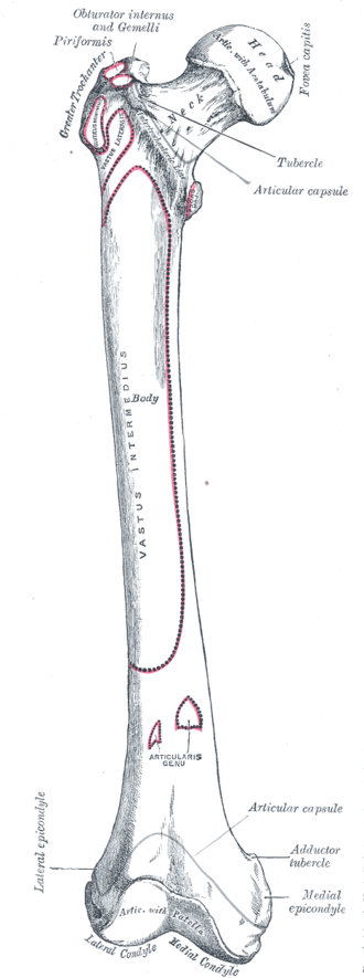
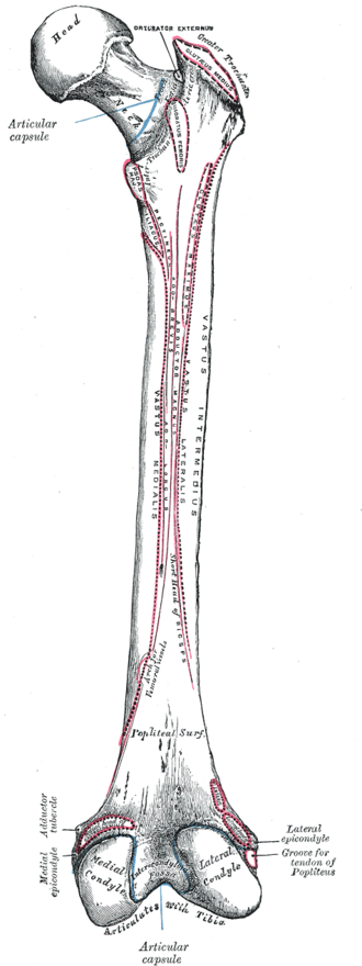
- Head: about two-thirds of a sphere, articulating with the acetabulum; the fovea gives attachment to the ligament of the head
- Neck: joins the head to the shaft at an angle of about 125° (angle of inclination) and is anteverted about 10–15°; a common fracture site
- Greater trochanter: attachment of gluteus medius and minimus, piriformis and the short lateral rotators; palpable on the lateral hip
- Lesser trochanter: attachment of iliopsoas
- Intertrochanteric line (anterior; capsule and iliofemoral ligament) and intertrochanteric crest (posterior; quadratus femoris)
- Shaft: bowed anteriorly; the linea aspera on its back gives attachment to the adductors, the short head of biceps and the vastus muscles; the gluteal tuberosity for gluteus maximus
- Distal end: medial and lateral condyles and epicondyles, the adductor tubercle (adductor magnus), the patellar surface and the intercondylar fossa (cruciate ligaments)
- Relations
The femoral vessels lie in front of the head in the femoral triangle. The sciatic nerve runs behind the hip, and the femoral artery winds around the shaft to become the popliteal artery behind the distal femur, where it is at risk in supracondylar fractures.
- Ossification
Primary center in the shaft appears in the 7th fetal week. The distal femoral epiphysis is usually present at birth (a sign of a full-term fetus). Secondary centers for the head (first year), greater trochanter and lesser trochanter fuse in late adolescence; the distal epiphysis fuses last (about 18–20 years). verify
- Notes
Blood supply of the head: mainly the medial circumflex femoral artery (retinacular branches running up the neck), with a small contribution from the artery of the ligament of the head (obturator). Displaced intracapsular neck fractures tear the retinacular vessels → avascular necrosis and nonunion.
Muscle attachments
- Adductor muscles of the thigh — insertion
- Gluteus maximus — insertion
- Gluteus medius and minimus — insertion
- Hamstrings — origin
- Piriformis — insertion
- Quadriceps femoris — origin
- Triceps surae and calcaneal tendon — origin
Ligament attachments
Joints
Mentioned in
Generated automatically from links in other entries.


Clinical correlations
Femoral neck (hip) fracture PANCE
- Mechanism / lesion
A low-energy fall in an older adult with osteoporosis; high-energy trauma in the young.
- Classic presentation
Hip or groin pain and inability to bear weight; the leg is shortened and externally rotated (displaced fractures). Intracapsular (neck) fractures risk AVN: displaced ones in older adults are usually treated with arthroplasty; intertrochanteric (extracapsular) fractures keep their blood supply and are fixed with a nail or sliding hip screw.
- Notes
Occult fractures may not show on the first X-ray → MRI. Surgery within about 24–48 hours reduces mortality.
Slipped capital femoral epiphysis (SCFE) PANCE
- Mechanism / lesion
The femoral head slips posteriorly and inferiorly on the neck through the growth plate; typically an obese adolescent boy (10–16 years), sometimes with endocrine disorders (hypothyroidism).
- Classic presentation
Hip, thigh or knee pain and a limp; the hip externally rotates on flexion, with limited internal rotation. Frog-leg lateral X-ray; treat with non-weight-bearing and urgent in situ screw fixation.
Legg–Calvé–Perthes disease and avascular necrosis PANCE
- Mechanism / lesion
Perthes: idiopathic AVN of the femoral head in children about 4–8 years (boys). Adult AVN: steroids, alcohol, sickle cell disease, SLE, trauma, caisson disease.
- Classic presentation
Insidious limp and hip/groin or knee pain with reduced internal rotation and abduction. X-ray may be normal early — MRI is most sensitive.
Femoral shaft fracture
- Mechanism / lesion
High-energy trauma; in young children consider non-accidental injury.
- Classic presentation
Deformity, swelling and pain; blood loss can exceed 1 L. Look for associated hip and knee injuries and fat embolism syndrome (hypoxia, confusion, petechial rash).
High-yield PANCE pearls
- Older woman after a fall, shortened, externally rotated leg → hip fracture; normal X-ray but high suspicion → MRI.
- Obese adolescent with knee pain and limp → SCFE (hip exam, frog-leg lateral); knee pain can be referred hip pain.
- Child 4–8 with a limp and hip/knee pain → Legg–Calvé–Perthes.
- Femoral head blood supply = medial circumflex femoral artery → AVN after displaced neck fracture.
- Long-bone fracture + hypoxia + confusion + petechiae → fat embolism.
Imaging anatomy
- X-ray
AP pelvis and lateral hip for hip fractures (look for disruption of the trabecular pattern and Shenton line). Frog-leg lateral views for SCFE (Klein line fails to intersect the epiphysis) and Perthes. Full-length femur films for shaft fractures, including the hip and knee.
- CT
Characterizes complex fractures and guides surgical planning.
- MRI
Detects occult hip fractures, stress fractures of the neck and early AVN.
- Key views & landmarks
- AP pelvis + cross-table/frog-leg lateral hip
- MRI hip for occult fracture or AVN
Physical exam
- Surface landmarks
The greater trochanter is palpable on the lateral thigh about a hand's breadth below the iliac crest; the medial and lateral femoral condyles and epicondyles at the knee; the adductor tubercle above the medial condyle.
- Palpation
Greater trochanteric tenderness suggests trochanteric bursitis/gluteal tendinopathy rather than a joint problem.
- Strength / function testing
Inspect leg length and rotation; in a child with a limp, always examine the hip, including internal rotation.
Self-quiz
A 13-year-old obese boy has 3 weeks of left knee pain and a limp. The knee exam is normal. When his left hip is flexed, it rotates externally, and internal rotation is limited. What is the most likely diagnosis?
Correct answer: B
SCFE classically presents in obese adolescents with hip, thigh or referred knee pain. Obligatory external rotation on hip flexion and loss of internal rotation are typical. Confirm with AP and frog-leg lateral X-rays, and stop weight-bearing until it is pinned.
Show answer
B
SCFE classically presents in obese adolescents with hip, thigh or referred knee pain. Obligatory external rotation on hip flexion and loss of internal rotation are typical. Confirm with AP and frog-leg lateral X-rays, and stop weight-bearing until it is pinned.
A displaced femoral neck fracture threatens the blood supply of the femoral head mainly by damaging branches of which artery?
Correct answer: B
The medial circumflex femoral artery gives retinacular branches along the neck to the head. Displaced intracapsular fractures tear them, leading to avascular necrosis; the artery of the ligament of the head contributes little in adults.
Show answer
B
The medial circumflex femoral artery gives retinacular branches along the neck to the head. Displaced intracapsular fractures tear them, leading to avascular necrosis; the artery of the ligament of the head contributes little in adults.
A 78-year-old woman falls at home and cannot bear weight. Her right leg is shortened and externally rotated, but hip X-rays show no fracture. What is the best next step?
Correct answer: B
With strong clinical suspicion and normal films, MRI is the most sensitive test for an occult hip fracture; delayed diagnosis risks displacement and AVN.
Show answer
B
With strong clinical suspicion and normal films, MRI is the most sensitive test for an occult hip fracture; delayed diagnosis risks displacement and AVN.
A 6-year-old boy has had an insidious limp and knee pain for 6 weeks. Hip internal rotation and abduction are reduced, and hip radiographs are normal. What is the most likely diagnosis and the most sensitive test?
Correct answer: B
Legg–Calvé–Perthes disease is idiopathic AVN of the femoral head in children about 4–8 years, causing a limp and hip or referred knee pain with reduced internal rotation and abduction. Radiographs can be normal early, and MRI is most sensitive. SCFE affects obese adolescents.
Show answer
B
Legg–Calvé–Perthes disease is idiopathic AVN of the femoral head in children about 4–8 years, causing a limp and hip or referred knee pain with reduced internal rotation and abduction. Radiographs can be normal early, and MRI is most sensitive. SCFE affects obese adolescents.
Two days after a femoral shaft fracture, a 22-year-old becomes hypoxic and confused and develops a petechial rash over the chest and axillae. What is the most likely diagnosis?
Correct answer: D
Fat embolism syndrome follows long-bone (especially femoral) fractures and classically causes hypoxia, confusion and a petechial rash, usually 1–3 days after injury.
Show answer
D
Fat embolism syndrome follows long-bone (especially femoral) fractures and classically causes hypoxia, confusion and a petechial rash, usually 1–3 days after injury.