Ischioanal fossa
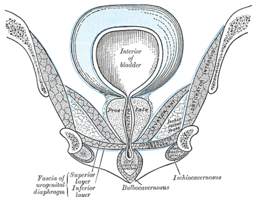
Paired fat-filled wedge-shaped spaces on either side of the anal canal. The pudendal canal lies in their lateral wall, and anorectal abscesses spread through them, sometimes in a horseshoe pattern.
Core anatomy
- Location
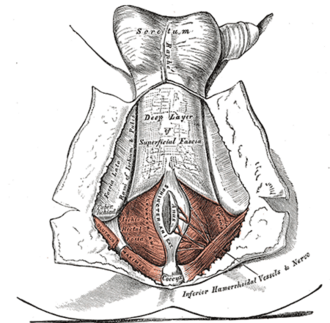
In the anal triangle of the perineum, between the anal canal and the ischium on each side.
- Boundaries
- lateral
- Ischium and obturator internus with its fascia (containing the pudendal canal)
- medial
- External anal sphincter and the sloping levator ani
- apex (superior)
- Where levator ani arises from the obturator fascia
- floor (inferior)
- Perianal skin and superficial fascia
- anterior
- Posterior edge of the perineal membrane (the anterior recesses extend forward above it)
- posterior
- Sacrotuberous ligament and gluteus maximus
- Contents
- Fat (the fat body of the ischioanal fossa), which lets the anal canal distend during defecation
- Pudendal canal in the lateral wall, containing the pudendal nerve and internal pudendal vessels
- Inferior rectal nerves and vessels, crossing from the pudendal canal medially to the anal canal
- Perineal branches of the posterior femoral cutaneous nerve and the perforating cutaneous nerve (S2–S3)
- Communications
The right and left fossae communicate behind the anal canal (deep postanal space, above the anococcygeal body), so infection can spread from one side to the other (horseshoe abscess). Anterior recesses extend forward above the perineal membrane.
Found in
Mentioned in
Generated automatically from links in other entries.
Swipe sideways to see the whole diagram →


Clinical correlations
Ischioanal (ischiorectal) abscess PANCE
- Mechanism / lesion
Spread of a cryptoglandular infection from an anal gland through the external sphincter into the fossa (or from a perianal abscess).
- Classic presentation
Deep throbbing buttock/perianal pain, fever, and a tender indurated or fluctuant area lateral to the anus (external signs may be subtle because the fossa is deep). Treat with prompt incision and drainage; antibiotics are added for cellulitis, immunosuppression or diabetes.
Fistula-in-ano PANCE
- Mechanism / lesion
A persistent tract between an anal gland at the pectinate line and the perianal skin after an abscess; associated with Crohn disease when complex or multiple.
- Classic presentation
Chronic intermittent discharge from a perianal opening. Goodsall rule: an external opening anterior to a transverse line through the anus usually has a straight radial tract; one posterior to it usually curves to the posterior midline. Treated surgically (fistulotomy, seton), taking care to preserve continence.
High-yield PANCE pearls
- Perianal pain + fever + fluctuance → incision and drainage (antibiotics alone are not enough).
- Goodsall rule: anterior openings → straight tracts; posterior openings → curve to the posterior midline.
- Horseshoe abscess = spread between the two ischioanal fossae behind the anal canal.
- Complex or recurrent fistulas → think Crohn disease.
Imaging anatomy
- CT
CT pelvis locates deep abscesses (ischioanal, supralevator).
- MRI
MRI (fistula protocol) is the gold standard for mapping complex fistula tracts and horseshoe extensions.
- Ultrasound
Endoanal ultrasound (with hydrogen peroxide enhancement) shows fistula tracts relative to the sphincters.
- Key views & landmarks
- MRI pelvis fistula protocol (axial and coronal STIR/T2)
Physical exam
- Surface landmarks
The fossa lies between the anus and the ischial tuberosity on each side.
- Palpation
Inspection and palpation of the buttock and perianal skin for induration and fluctuance; DRE for tenderness or a mass lateral to the anal canal and for an internal opening.
Self-quiz
A 40-year-old man with diabetes has 4 days of throbbing left buttock pain and fever. There is a tender, indurated, fluctuant area 3 cm lateral to the anus. What is the most appropriate management?
Correct answer: B
This is an ischioanal abscess. Definitive treatment is incision and drainage; antibiotics are an adjunct in patients with diabetes, immunosuppression or cellulitis.
Show answer
B
This is an ischioanal abscess. Definitive treatment is incision and drainage; antibiotics are an adjunct in patients with diabetes, immunosuppression or cellulitis.
Which structure runs in the lateral wall of the ischioanal fossa?
Correct answer: B
The pudendal (Alcock) canal is a split in the obturator internus fascia on the lateral wall of the ischioanal fossa; it carries the pudendal nerve and internal pudendal vessels.
Show answer
B
The pudendal (Alcock) canal is a split in the obturator internus fascia on the lateral wall of the ischioanal fossa; it carries the pudendal nerve and internal pudendal vessels.
How can an abscess in one ischioanal fossa spread to the other side?
Correct answer: A
The two ischioanal fossae communicate behind the anal canal through the deep postanal space, above the anococcygeal body, so infection can spread from side to side — a horseshoe abscess.
Show answer
A
The two ischioanal fossae communicate behind the anal canal through the deep postanal space, above the anococcygeal body, so infection can spread from side to side — a horseshoe abscess.