Collateral ligaments of the knee
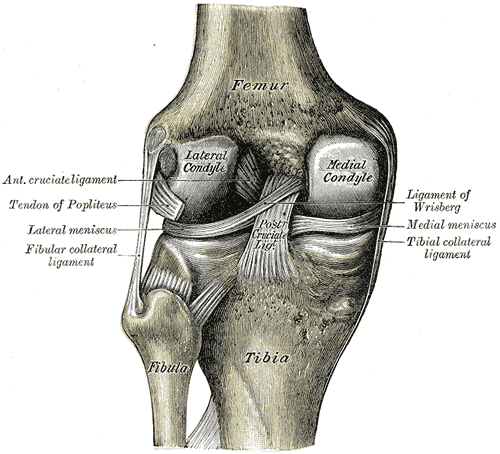
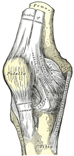
Side ligaments of the knee — the broad MCL, attached to the medial meniscus, resists valgus stress; the cord-like LCL resists varus stress. The MCL is the most commonly injured knee ligament.
Core anatomy
- Location
On the medial and lateral sides of the knee joint.
- Attachments
MCL (tibial collateral): a flat band from the medial epicondyle of the femur to the medial condyle and upper medial shaft of the tibia; its deep fibers are firmly attached to the medial meniscus and capsule. LCL (fibular collateral): a round cord from the lateral epicondyle to the head of the fibula, separate from the capsule and lateral meniscus (the popliteus tendon passes between them).
- Function
MCL resists valgus (abduction) force and external rotation; LCL resists varus (adduction) force. Both are taut in extension.
- Relations
The pes anserinus (sartorius, gracilis, semitendinosus) crosses superficial to the distal MCL. The common fibular nerve lies close to the LCL and fibular head — assess it with lateral/posterolateral injuries.
Swipe sideways to see the whole diagram →


Clinical correlations
MCL sprain PANCE
- Mechanism / lesion
A valgus blow to the lateral knee (e.g. a football tackle) or valgus twisting.
- Classic presentation
Medial knee pain and tenderness along the ligament; pain or laxity on valgus stress at 30° of flexion. Graded I–III. Isolated MCL injuries heal well non-operatively (hinged brace and rehab). Look for associated ACL and medial meniscal tears (unhappy triad).
LCL and posterolateral corner injury
- Mechanism / lesion
A varus blow to the medial knee or hyperextension–varus injury; often combined with cruciate injury.
- Classic presentation
Lateral knee pain, varus laxity; check the common fibular nerve (foot drop) and posterolateral corner structures; often needs surgical repair.
Pes anserine bursitis
- Mechanism / lesion
Irritation of the bursa beneath the pes anserinus, common with knee OA and obesity.
- Classic presentation
Tenderness about 2–5 cm below the medial joint line over the proximal medial tibia, below the MCL's joint-line portion.
High-yield PANCE pearls
- Valgus blow → MCL; varus blow → LCL.
- Test collaterals at 30° of flexion (to isolate them from the cruciates and capsule).
- Isolated MCL tear → brace and rehab (non-operative).
- LCL/posterolateral injury → check the common fibular nerve.
Imaging anatomy
- X-ray
Rules out fractures; the Pellegrini–Stieda lesion (calcification at the medial femoral epicondyle) indicates a chronic MCL injury.
- MRI
Grades collateral injuries and detects associated cruciate, meniscal and posterolateral corner injuries.
- Ultrasound
Dynamic assessment of the MCL.
Physical exam
- Surface landmarks
The MCL runs from the medial epicondyle across the joint line to the upper medial tibia; the LCL is palpable as a cord between the lateral epicondyle and fibular head when the leg is crossed in "figure-4".
- Palpation
Tenderness along the course of each ligament and at its attachments.
Valgus stress test
- Technique
Apply a valgus (abduction) force at the knee while stabilizing the ankle, at 0° and at 30° of flexion.
- Positive test
Pain or medial joint-line gapping. Laxity at 30° = isolated MCL; laxity also at 0° = additional cruciate/capsular injury.
- Significance
MCL injury.
Varus stress test
- Technique
Apply a varus (adduction) force at 0° and 30° of flexion.
- Positive test
Pain or lateral gapping.
- Significance
LCL (and, at 0°, posterolateral corner/cruciate) injury.
Self-quiz
A football player is tackled from the side, with a blow to the lateral aspect of his planted knee. He has medial knee pain. Which structure is most likely injured?
Correct answer: B
A blow to the lateral knee produces a valgus force that stretches the medial side, injuring the MCL (often with the ACL and medial meniscus).
Show answer
B
A blow to the lateral knee produces a valgus force that stretches the medial side, injuring the MCL (often with the ACL and medial meniscus).
Why is the medial meniscus injured more often than the lateral meniscus?
Correct answer: B
The medial meniscus is anchored to the MCL and capsule, so it moves less and is trapped more easily. The LCL is separate from the lateral meniscus, which is also partly mobilized by popliteus.
Show answer
B
The medial meniscus is anchored to the MCL and capsule, so it moves less and is trapped more easily. The LCL is separate from the lateral meniscus, which is also partly mobilized by popliteus.
Why are the collateral ligaments of the knee tested with the knee flexed to about 30°?
Correct answer: A
At 30° of flexion, the cruciates and posterior capsule contribute less to varus/valgus stability, so laxity reflects the collateral ligament itself. Both collaterals are taut in extension; laxity in full extension implies a more extensive injury.
Show answer
A
At 30° of flexion, the cruciates and posterior capsule contribute less to varus/valgus stability, so laxity reflects the collateral ligament itself. Both collaterals are taut in extension; laxity in full extension implies a more extensive injury.
A rugby player takes a blow to the medial side of his knee. He has lateral knee pain, varus laxity and a new foot drop. Which ligament and nerve are most likely injured?
Correct answer: B
A varus force (blow to the medial knee) injures the LCL and posterolateral corner. The common fibular nerve lies close to the LCL and fibular head, so check for foot drop. A valgus blow injures the MCL.
Show answer
B
A varus force (blow to the medial knee) injures the LCL and posterolateral corner. The common fibular nerve lies close to the LCL and fibular head, so check for foot drop. A valgus blow injures the MCL.
A 62-year-old woman with obesity and knee osteoarthritis has pain and tenderness over the proximal medial tibia, about 3 cm below the joint line. Valgus stress is painless. What is the most likely diagnosis?
Correct answer: E
Pes anserine bursitis causes tenderness about 2–5 cm below the medial joint line over the proximal medial tibia, where sartorius, gracilis and semitendinosus insert superficial to the distal MCL. It is common with knee OA and obesity. MCL sprain hurts with valgus stress; meniscal tears cause joint-line tenderness.
Show answer
E
Pes anserine bursitis causes tenderness about 2–5 cm below the medial joint line over the proximal medial tibia, where sartorius, gracilis and semitendinosus insert superficial to the distal MCL. It is common with knee OA and obesity. MCL sprain hurts with valgus stress; meniscal tears cause joint-line tenderness.
Flashcards
See also
- Knee joint
- Cruciate ligaments
- Menisci of the knee
- Knee ligament and meniscal injuries (comparison table)
- A pop in the knee on landing (clinical case)