Pleural cavity
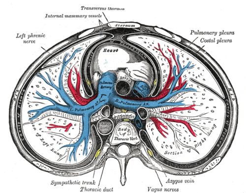
Potential space between the visceral and parietal pleura around each lung. It's the site of pneumothorax and pleural effusion and the target of thoracentesis and chest tubes.
Core anatomy
- Location
Each hemithorax, surrounding a lung. The two cavities are completely separate.
- Boundaries
- visceral pleura
- Adheres to the lung surface and into the fissures. Insensitive to pain (autonomic innervation only)
- costal parietal pleura
- Lines the internal thoracic wall. Somatic sensation via the intercostal nerves (localized pain)
- diaphragmatic parietal pleura
- Covers the diaphragm. Central part via the phrenic nerve (referred shoulder pain); peripheral part via the intercostal nerves
- mediastinal parietal pleura
- Covers the mediastinum. Sensation via the phrenic nerve
- cervical pleura (cupula)
- Extends 2–3 cm above the medial third of the clavicle into the root of the neck
- Contents
- A thin film of serous (pleural) fluid that lubricates the surfaces and couples the lung to the chest wall
- Communications
The visceral and parietal layers are continuous around the root of the lung, where a fold hangs below it (the pulmonary ligament). Recesses, not filled by lung except in deep inspiration: the costodiaphragmatic recess (lowest point, where fluid collects) and the costomediastinal recess (anterior, behind the sternum).
- Notes
Surface markings of the lower pleural reflection (about two ribs below the lower lung border): 8th rib at the midclavicular line, 10th rib at the mid-axillary line, 12th rib posteriorly near the vertebral column. The lung's lower border is at the 6th, 8th and 10th ribs at the same lines. The gap between them is the costodiaphragmatic recess. verify

Clinical correlations
Pneumothorax PANCE
- Mechanism / lesion
Air in the pleural cavity. Primary spontaneous: rupture of apical blebs, typically in tall, thin young men (and smokers). Secondary: underlying lung disease (COPD, cystic fibrosis). Traumatic or iatrogenic (central lines, thoracentesis, mechanical ventilation).
- Classic presentation
Sudden pleuritic chest pain and dyspnea. Decreased breath sounds, hyperresonance and decreased tactile fremitus on the affected side.
Tension pneumothorax PANCE
- Mechanism / lesion
A one-way valve lets air enter the pleural cavity but not leave. Rising pressure shifts the mediastinum and impairs venous return.
- Classic presentation
Respiratory distress, hypotension, distended neck veins, absent breath sounds and hyperresonance on the affected side, tracheal deviation away from the affected side (a late sign). It's a clinical diagnosis: treat with immediate needle decompression or finger thoracostomy, then a chest tube, without waiting for imaging.
Pleural effusion PANCE
- Mechanism / lesion
Excess fluid from increased hydrostatic or decreased oncotic pressure (transudate: heart failure, cirrhosis, nephrotic syndrome) or from inflammation or malignancy (exudate: pneumonia/parapneumonic, cancer, PE, TB). Light's criteria classify it using pleural/serum protein and LDH.
- Classic presentation
Dyspnea and pleuritic pain. Dullness to percussion, decreased breath sounds and decreased tactile fremitus over the fluid, sometimes with egophony at its upper border.
Hemothorax, empyema and chylothorax
- Mechanism / lesion
Blood (trauma, intercostal or internal thoracic artery injury), pus (complicated pneumonia), or lymph (injury to the thoracic duct) in the pleural cavity.
- Classic presentation
Effusion signs plus the context. Diagnosed by fluid analysis (hematocrit, pH/glucose/Gram stain, triglycerides).
High-yield PANCE pearls
- Tension pneumothorax: hypotension + JVD + absent breath sounds + tracheal deviation away → needle decompression now.
- Effusion: dull, ↓ fremitus. Pneumothorax: hyperresonant, ↓ fremitus. Consolidation: dull, ↑ fremitus.
- Primary spontaneous pneumothorax: tall, thin young male, apical blebs.
- Light's criteria (exudate if any): pleural/serum protein > 0.5, pleural/serum LDH > 0.6, pleural LDH > 2/3 of the upper limit of normal serum LDH.
- Parietal pleura is pain-sensitive; visceral pleura is not.
Imaging anatomy
- X-ray
Pneumothorax: a thin visceral pleural line with no lung markings beyond it (best on an upright, ideally expiratory, film). On a supine film look for the deep sulcus sign. Effusion: blunting of the costophrenic angle and a meniscus. A lateral film detects smaller volumes than a PA film (roughly 50 mL vs 200 mL). verify Large effusions shift the mediastinum away; large volume loss pulls it toward.
- CT
The most sensitive test for small pneumothoraces and for characterizing effusions (loculation, empyema with the "split pleura" sign, pleural masses).
- Ultrasound
Effusion: an anechoic space above the diaphragm; the "spine sign" (vertebral bodies visible above the diaphragm). Ultrasound detects smaller effusions than CXR and guides thoracentesis. Pneumothorax: absent lung sliding, absent B-lines, "barcode/stratosphere" sign on M-mode, and a lung point (specific for pneumothorax). Lung sliding makes pneumothorax unlikely at that spot.
- Key views & landmarks
- Upright PA and lateral CXR (expiratory film for small pneumothorax)
- Point-of-care ultrasound: anterior chest (pneumothorax), posterolateral bases (effusion)
Physical exam
- Surface landmarks
Needle decompression: 2nd intercostal space in the midclavicular line, or the 4th/5th intercostal space in the anterior to mid-axillary line (current ATLS guidance for adults favors the latter). Chest tube "triangle of safety": lateral border of pectoralis major, anterior border of latissimus dorsi, a horizontal line at the level of the nipple (about the 5th intercostal space), apex below the axilla. Thoracentesis: posteriorly, about one or two interspaces below the top of the effusion (ultrasound-guided), above the diaphragm, and over the superior border of the rib.
- Palpation
Tactile fremitus ("99") is decreased over effusion and pneumothorax and increased over consolidation. Check tracheal position in the suprasternal notch.
- Strength / function testing
Percussion (dull vs hyperresonant), auscultation (breath sounds, friction rub, egophony at the top of an effusion).
Self-quiz
A 24-year-old man is stabbed in the right chest. He is hypotensive with distended neck veins, absent breath sounds on the right, and hyperresonance to percussion. What is the next step?
Correct answer: C
This is tension pneumothorax, a clinical diagnosis. Decompress immediately (needle or finger thoracostomy), then place a chest tube. Waiting for imaging delays treatment. Pericardiocentesis treats tamponade, which also causes JVD and hypotension but with muffled heart sounds and equal breath sounds.
Show answer
C
This is tension pneumothorax, a clinical diagnosis. Decompress immediately (needle or finger thoracostomy), then place a chest tube. Waiting for imaging delays treatment. Pericardiocentesis treats tamponade, which also causes JVD and hypotension but with muffled heart sounds and equal breath sounds.
Examination of the left lung base shows dullness to percussion, decreased breath sounds and decreased tactile fremitus. What is the most likely finding?
Correct answer: B
Dullness with decreased fremitus = pleural effusion (fluid separates the lung from the chest wall). Consolidation is dull with increased fremitus. Pneumothorax is hyperresonant.
Show answer
B
Dullness with decreased fremitus = pleural effusion (fluid separates the lung from the chest wall). Consolidation is dull with increased fremitus. Pneumothorax is hyperresonant.
Where is the needle inserted during thoracentesis to avoid the intercostal neurovascular bundle?
Correct answer: B
The bundle runs in the costal groove along the inferior border of each rib, so the needle passes just over the superior border of the rib below.
Show answer
B
The bundle runs in the costal groove along the inferior border of each rib, so the needle passes just over the superior border of the rib below.
On a point-of-care ultrasound of the anterior chest, which finding is most specific for pneumothorax?
Correct answer: C
The lung point (where sliding lung meets the non-sliding pneumothorax) is highly specific. Lung sliding and B-lines rule pneumothorax out at that location. Anechoic fluid and the spine sign indicate effusion.
Show answer
C
The lung point (where sliding lung meets the non-sliding pneumothorax) is highly specific. Lung sliding and B-lines rule pneumothorax out at that location. Anechoic fluid and the spine sign indicate effusion.
In an upright patient, where does a small pleural effusion collect first?
Correct answer: E
The costodiaphragmatic recess is the lowest part of the pleural cavity, where fluid collects first (blunting of the costophrenic angle on CXR). The costomediastinal recess lies anteriorly behind the sternum.
Show answer
E
The costodiaphragmatic recess is the lowest part of the pleural cavity, where fluid collects first (blunting of the costophrenic angle on CXR). The costomediastinal recess lies anteriorly behind the sternum.
Flashcards
See also
- Lungs
- Intercostal space
- Stab wound to the chest (clinical case)