Submandibular gland
Salivary gland under the body of the mandible that produces most resting saliva. It is the most common site of salivary stones because its duct runs uphill and its saliva is thick.
Core anatomy
- Location
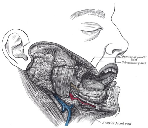
In the submandibular triangle (see triangles of the neck), wrapped around the posterior border of mylohyoid, with a large superficial part and a small deep part.
- Parts
- Superficial part: below mylohyoid, in the submandibular triangle
- Deep part: above mylohyoid, in the floor of the mouth
- Submandibular (Wharton) duct: runs forward in the floor of the mouth to open at the sublingual papilla beside the frenulum of the tongue; the lingual nerve loops under it
- Relations
The marginal mandibular branch of the facial nerve, the lingual nerve and the hypoglossal nerve are all close — at risk in submandibular gland excision.
- Arterial supply
Facial artery (from the external carotid artery), which grooves the gland.
- Venous drainage
Facial vein.
- Lymphatic drainage
Submandibular nodes (see cervical lymph nodes).
- Innervation
Parasympathetic secretomotor: facial nerve (CN VII) — superior salivatory nucleus → chorda tympani (joins the lingual nerve) → submandibular ganglion.

Clinical correlations
Sialolithiasis (salivary stones) PANCE
- Mechanism / lesion
About 80–90% of salivary stones occur in the submandibular gland — its saliva is more mucous and alkaline and its duct is long and runs upward. verify
- Classic presentation
Painful swelling under the jaw at mealtimes that subsides afterwards; a stone may be palpable in the floor of the mouth. Treat with hydration, sialagogues (lemon drops), massage and warm compresses; removal or sialendoscopy if persistent.
Sialadenitis
- Mechanism / lesion
Bacterial infection behind an obstructing stone.
- Classic presentation
Painful, red, tender swelling with pus from the duct; antibiotics and treat the stone.
Ludwig angina PANCE
- Mechanism / lesion
Rapidly spreading cellulitis of the submandibular and sublingual spaces, usually from a lower molar infection.
- Classic presentation
Brawny swelling of the floor of the mouth, tongue elevation, drooling and airway compromise — secure the airway and give IV antibiotics. See deep neck spaces.
High-yield PANCE pearls
- Swelling under the jaw at mealtimes → submandibular stone (most common site).
- Wharton duct opens at the sublingual papilla; Stensen duct opposite the upper 2nd molar.
- Submandibular secretomotor = CN VII (chorda tympani).
Imaging anatomy
- CT
Non-contrast CT detects stones; contrast CT for abscess and Ludwig angina.
- Ultrasound
First-line for stones and gland swelling.
Physical exam
- Surface landmarks
Below the body of the mandible, in front of the angle.
- Palpation
Bimanual palpation — one finger in the floor of the mouth, the other under the jaw — feels the gland and stones along the duct; inspect the duct openings for pus.
Self-quiz
Why are salivary stones most common in the submandibular gland?
Correct answer: B
The submandibular gland produces more mucous, alkaline, calcium-rich saliva, and its long Wharton duct runs uphill against gravity, favoring stone formation.
Show answer
B
The submandibular gland produces more mucous, alkaline, calcium-rich saliva, and its long Wharton duct runs uphill against gravity, favoring stone formation.
A 40-year-old man has swelling and pain under the left side of his jaw every time he eats, which resolves an hour later. What is the most likely diagnosis?
Correct answer: B
Meal-related swelling and pain that subsides is classic for a stone obstructing the duct (sialolithiasis), most often in the submandibular gland.
Show answer
B
Meal-related swelling and pain that subsides is classic for a stone obstructing the duct (sialolithiasis), most often in the submandibular gland.
Where does the submandibular (Wharton) duct open?
Correct answer: B
The Wharton duct runs forward in the floor of the mouth, with the lingual nerve looping under it, to open at the sublingual papilla. The parotid (Stensen) duct opens opposite the upper second molar.
Show answer
B
The Wharton duct runs forward in the floor of the mouth, with the lingual nerve looping under it, to open at the sublingual papilla. The parotid (Stensen) duct opens opposite the upper second molar.