Pons
Middle part of the brainstem that links the cerebrum to the cerebellum. It holds the nuclei of CN V–VIII and horizontal gaze centers; basilar artery occlusion causes locked-in syndrome.
Core anatomy
- Location
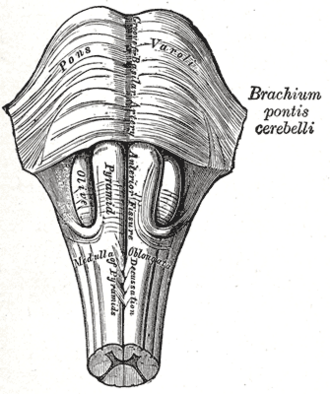
Between the midbrain and the medulla oblongata, in front of the cerebellum and the fourth ventricle, resting on the clivus.
- Parts
- Basilar (ventral) pons: descending corticospinal and corticobulbar fibers, pontine nuclei, and transverse pontocerebellar fibers forming the middle cerebellar peduncle
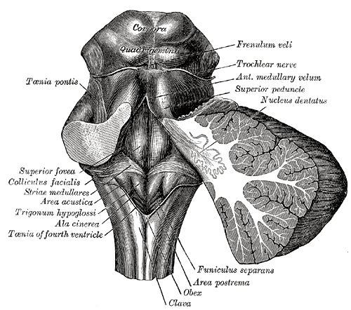
- Tegmentum (dorsal): nuclei of CN V (motor and principal sensory), VI, VII and VIII (vestibular and cochlear); the PPRF (paramedian pontine reticular formation — horizontal gaze center); MLF; medial lemniscus and spinothalamic tract; locus coeruleus (norepinephrine)
- Floor of the fourth ventricle with the facial colliculus (CN VII fibers looping around the CN VI nucleus)
- Respiratory centers (pneumotaxic and apneustic) that modulate medullary rhythm
- Relations
The basilar artery runs in the basilar groove on its anterior surface.
- Arterial supply
Basilar artery — paramedian and short circumferential pontine branches; AICA (lateral lower pons) and SCA (upper pons) — see vertebrobasilar system.
- Venous drainage
Pontine veins → superior petrosal sinus and basal veins.
- Notes
Emergence of CN V (lateral pons), CN VI (pontomedullary junction, medially), CN VII and VIII (cerebellopontine angle) — see trigeminal nerve (CN V), abducens nerve (CN VI), facial nerve (CN VII), vestibulocochlear nerve (CN VIII).
Mentioned in
- Abducens nerve (CN VI)
- Cerebellum
- Corticospinal tract
- Facial nerve (CN VII)
- Medulla oblongata
- Midbrain
- Trigeminal nerve (CN V)
- Vertebrobasilar system
Generated automatically from links in other entries.
Swipe sideways to see the whole diagram →


Clinical correlations
Locked-in syndrome PANCE
- Mechanism / lesion
Basilar artery occlusion (or hemorrhage) destroying the ventral pons — corticospinal and corticobulbar tracts — while sparing the tegmentum (reticular activating system) above.
- Classic presentation
Quadriplegia and anarthria with preserved consciousness; vertical eye movements and blinking are preserved (midbrain) and used to communicate. Sudden basilar occlusion can present with coma, and needs emergency thrombectomy.
Osmotic demyelination syndrome (central pontine myelinolysis) PANCE
- Mechanism / lesion
Overly rapid correction of chronic hyponatremia (risk: alcohol use disorder, malnutrition, liver disease, hypokalemia).
- Classic presentation
Days after correction: dysarthria, dysphagia, quadriparesis, locked-in state. Prevent by correcting sodium no faster than about 8 mEq/L per 24 hours in high-risk patients (10–12 limit otherwise).
Pontine hemorrhage and gaze palsies PANCE
- Mechanism / lesion
Hypertensive hemorrhage from paramedian perforators; lesions of the PPRF, abducens nucleus or MLF.
- Classic presentation
Pontine hemorrhage: coma, quadriplegia, pinpoint reactive pupils, hyperthermia. PPRF/abducens nucleus lesion → ipsilateral horizontal gaze palsy. MLF → internuclear ophthalmoplegia (MS). "One-and-a-half syndrome" → both.
High-yield PANCE pearls
- Awake, quadriplegic, communicates by vertical eye movements/blinking → locked-in syndrome (ventral pons, basilar).
- Rapid correction of hyponatremia → osmotic demyelination → ≤ 8 mEq/L/24 h in high-risk patients.
- Coma + pinpoint pupils → pontine hemorrhage (or opioids).
- Pons houses nuclei of CN V, VI, VII, VIII.
Imaging anatomy
- CT
CT shows pontine hemorrhage; small infarcts are often missed in the posterior fossa.
- MRI
MRI with DWI for pontine infarcts and demyelination (a "trident" or "bat-wing" T2 lesion in osmotic demyelination, which may appear only after 1–2 weeks); MRA/CTA for basilar occlusion.
Physical exam
- Strength / function testing
Corneal reflex, facial strength, horizontal gaze (PPRF, CN VI), hearing and vestibular function; limb strength and reflexes for corticospinal involvement; level of consciousness and pupil size.
Self-quiz
A 60-year-old man is found unable to move his limbs or speak. He is alert and answers yes/no questions by looking up and down, but cannot move his eyes horizontally. Which artery is most likely occluded?
Correct answer: B
Locked-in syndrome from basilar artery occlusion infarcts the ventral pons (motor tracts) and the horizontal gaze centers, sparing consciousness and midbrain-controlled vertical gaze and blinking.
Show answer
B
Locked-in syndrome from basilar artery occlusion infarcts the ventral pons (motor tracts) and the horizontal gaze centers, sparing consciousness and midbrain-controlled vertical gaze and blinking.
A woman with alcohol use disorder has a sodium of 108 mEq/L, which rises to 128 mEq/L in 24 hours. Four days later she develops dysarthria and weakness of all limbs. What is the diagnosis?
Correct answer: B
Correcting chronic hyponatremia by 20 mEq/L in 24 hours is far too fast and causes osmotic demyelination (central pontine myelinolysis) — dysarthria, dysphagia and quadriparesis days later.
Show answer
B
Correcting chronic hyponatremia by 20 mEq/L in 24 hours is far too fast and causes osmotic demyelination (central pontine myelinolysis) — dysarthria, dysphagia and quadriparesis days later.
A comatose patient has pinpoint but reactive pupils and quadriplegia after sudden collapse. Where is the likely lesion (if opioids are excluded)?
Correct answer: B
Pontine hemorrhage causes coma, quadriplegia and pinpoint reactive pupils (interrupted descending sympathetic fibers). Opioid overdose is the main mimic.
Show answer
B
Pontine hemorrhage causes coma, quadriplegia and pinpoint reactive pupils (interrupted descending sympathetic fibers). Opioid overdose is the main mimic.
The locus coeruleus in the pons is the main source of which neurotransmitter?
Correct answer: D
The locus coeruleus is the brain's main source of norepinephrine. The substantia nigra supplies dopamine and the raphe nuclei serotonin.
Show answer
D
The locus coeruleus is the brain's main source of norepinephrine. The substantia nigra supplies dopamine and the raphe nuclei serotonin.
What is the function of the paramedian pontine reticular formation (PPRF)?
Correct answer: A
The PPRF is the horizontal gaze center, driving the ipsilateral abducens nucleus. Vertical gaze is controlled in the midbrain.
Show answer
A
The PPRF is the horizontal gaze center, driving the ipsilateral abducens nucleus. Vertical gaze is controlled in the midbrain.