Mitral valve
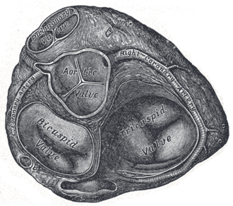
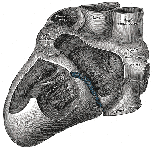
Two-leaflet valve between the left atrium and left ventricle, anchored by chordae tendineae to two papillary muscles. Mitral stenosis, regurgitation and prolapse are heard best at the apex.
Core anatomy
- Location
Between the left atrium and left ventricle, posterior to the sternum at about the level of the left 4th costal cartilage. verify
- Parts
- Anterior (aortic) leaflet and posterior (mural) leaflet
- Chordae tendineae attach the leaflets to the anterolateral and posteromedial papillary muscles, preventing prolapse into the atrium during systole
- Annulus: part of the fibrous skeleton of the heart
- Relations
The anterior leaflet is continuous with the aortic valve. The left atrium lies behind it, directly in front of the esophagus. The circumflex artery and coronary sinus run in the adjacent AV groove.
- Arterial supply
Papillary muscles: the anterolateral muscle usually has a dual supply (LAD and circumflex); the posteromedial muscle usually has a single supply (the posterior interventricular artery, from the RCA in right dominance), so it's more vulnerable to rupture in inferior MI.
- Notes
Auscultation area: the apex, left 5th intercostal space in the midclavicular line; the left lateral decubitus position accentuates mitral murmurs (especially stenosis, with the bell).


Clinical correlations
Mitral stenosis PANCE
- Mechanism / lesion
Almost always from rheumatic heart disease (commissural fusion). Rarely from annular calcification.
- Classic presentation
Dyspnea, orthopnea, hemoptysis, and atrial fibrillation (with risk of LA thrombus and stroke). A loud S1, an opening snap, then a low-pitched mid-diastolic rumble at the apex. A shorter S2–opening snap interval means more severe stenosis. The enlarged left atrium can cause dysphagia or hoarseness (Ortner syndrome).
Mitral regurgitation PANCE
- Mechanism / lesion
Primary: mitral valve prolapse, endocarditis, rheumatic disease. Secondary (functional): LV dilation stretching the annulus. Acute: papillary muscle rupture after MI (typically about 2–7 days after, posteromedial muscle, inferior MI) or chordal rupture.
- Classic presentation
Holosystolic, blowing murmur at the apex radiating to the axilla. Acute severe MR: sudden pulmonary edema and cardiogenic shock (the murmur may be soft).
Mitral valve prolapse PANCE
- Mechanism / lesion
Myxomatous degeneration with billowing of the leaflets into the left atrium in systole. Associated with Marfan and Ehlers–Danlos syndromes.
- Classic presentation
Mid-systolic click followed by a late systolic murmur. Maneuvers that decrease preload (standing, Valsalva) make the click earlier and the murmur longer; squatting has the opposite effect.
High-yield PANCE pearls
- MS: opening snap + mid-diastolic rumble at the apex; rheumatic; AFib; LA enlargement.
- MR: holosystolic murmur at the apex radiating to the axilla.
- MVP: mid-systolic click; standing/Valsalva → earlier click, longer murmur.
- Post-MI new holosystolic murmur + pulmonary edema → papillary muscle rupture (posteromedial, inferior MI).
Imaging anatomy
- X-ray
MS: LA enlargement (straightened left heart border, double density, splayed carina), pulmonary venous hypertension (upper-lobe vascular redistribution, Kerley B lines).
- MRI
Quantifies MR severity and LV volumes when echo is inconclusive.
- Ultrasound
Echocardiography: MS shows thickened leaflets with commissural fusion and doming of the anterior leaflet ("hockey stick" appearance), and valve area by planimetry or pressure half-time. MR shows the regurgitant jet (color Doppler) and its mechanism (prolapse, flail leaflet, ruptured chordae or papillary muscle). TEE is best for detailed mitral anatomy and LA appendage thrombus.
- Key views & landmarks
- Parasternal long axis, apical 4-chamber (color Doppler)
- TEE for surgical/percutaneous planning and LA appendage thrombus
Physical exam
- Surface landmarks
Mitral area: the apex, left 5th intercostal space in the midclavicular line.
- Palpation
Laterally displaced, diffuse apical impulse in chronic MR (LV dilation). A tapping apex (palpable S1) in MS.
- Strength / function testing
Listen at the apex with the bell in the left lateral decubitus position for the MS rumble. Use the diaphragm for the MR murmur and follow it toward the axilla. Stand and squat the patient to evaluate MVP.
Self-quiz
A 45-year-old woman who immigrated from a country with limited healthcare has dyspnea and palpitations. An ECG shows atrial fibrillation. There is an opening snap followed by a low-pitched diastolic rumble at the apex. What is the most likely underlying cause?
Correct answer: B
Mitral stenosis (opening snap and diastolic rumble) is almost always caused by rheumatic heart disease. The enlarged left atrium predisposes to atrial fibrillation.
Show answer
B
Mitral stenosis (opening snap and diastolic rumble) is almost always caused by rheumatic heart disease. The enlarged left atrium predisposes to atrial fibrillation.
Four days after an inferior STEMI, a patient develops sudden dyspnea, pulmonary edema and a new holosystolic murmur at the apex. Which structure most likely failed?
Correct answer: B
The posteromedial papillary muscle usually has a single blood supply (the posterior interventricular artery, from the RCA in right dominance), so it's more likely to rupture after an inferior MI, causing acute MR.
Show answer
B
The posteromedial papillary muscle usually has a single blood supply (the posterior interventricular artery, from the RCA in right dominance), so it's more likely to rupture after an inferior MI, causing acute MR.
A young woman has a mid-systolic click followed by a late systolic murmur. What happens to the click when she stands up from squatting?
Correct answer: B
Standing decreases preload (smaller LV volume), so the prolapse occurs earlier, moving the click earlier and lengthening the murmur.
Show answer
B
Standing decreases preload (smaller LV volume), so the prolapse occurs earlier, moving the click earlier and lengthening the murmur.
Which papillary muscle of the left ventricle usually has a dual blood supply from the LAD and circumflex?
Correct answer: A
The anterolateral papillary muscle usually has a dual supply (LAD and circumflex). The posteromedial muscle usually depends on a single artery (the posterior interventricular), so it is more vulnerable to rupture after inferior MI.
Show answer
A
The anterolateral papillary muscle usually has a dual supply (LAD and circumflex). The posteromedial muscle usually depends on a single artery (the posterior interventricular), so it is more vulnerable to rupture after inferior MI.
Which murmur is characteristic of chronic mitral regurgitation?
Correct answer: B
MR causes a holosystolic, blowing murmur at the apex radiating to the axilla. The other choices describe aortic stenosis, aortic regurgitation, mitral stenosis and tricuspid regurgitation.
Show answer
B
MR causes a holosystolic, blowing murmur at the apex radiating to the axilla. The other choices describe aortic stenosis, aortic regurgitation, mitral stenosis and tricuspid regurgitation.
Flashcards
See also
- Aortic valve
- Heart
- Chest pain with a slow pulse (clinical case)